

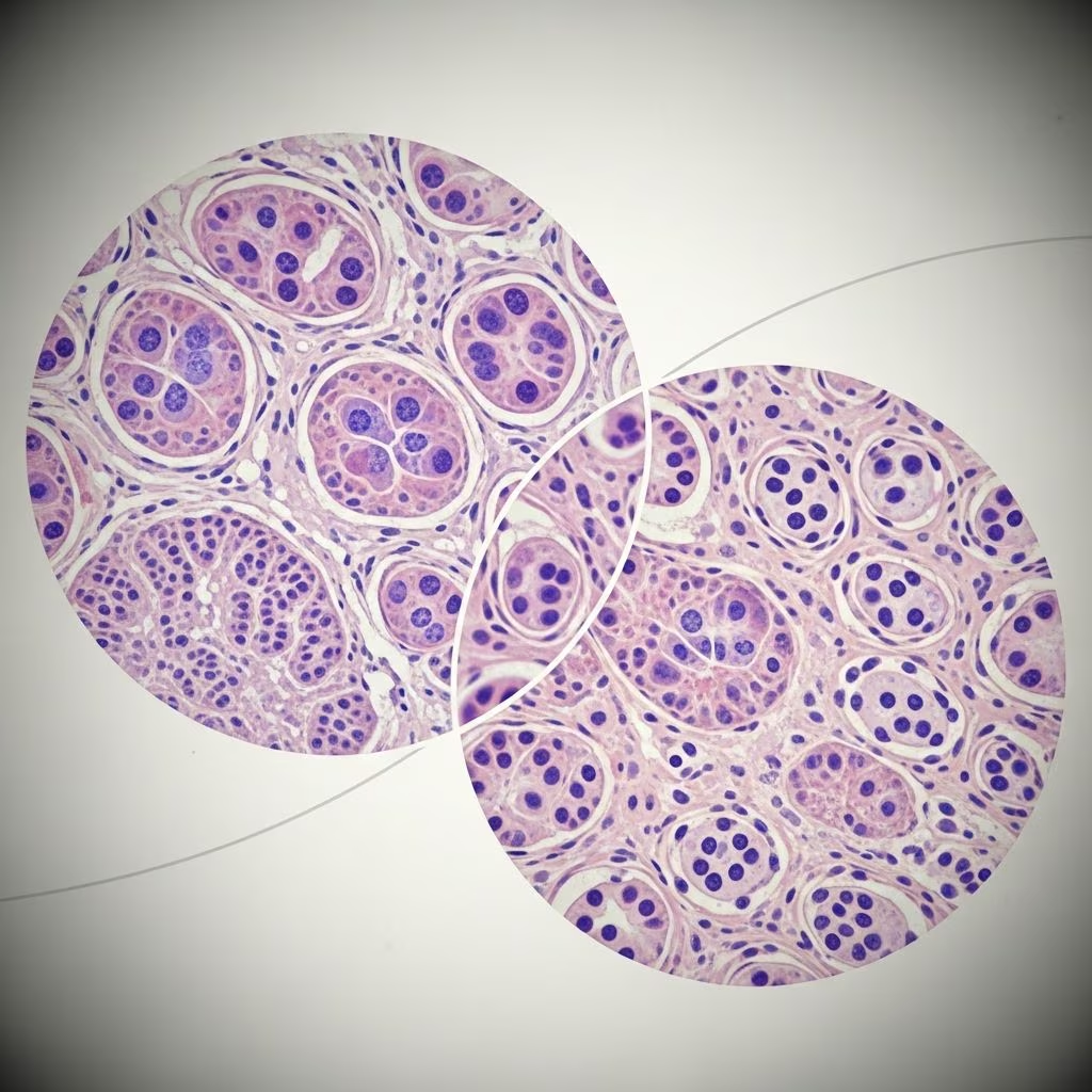
Understanding Epithelial Liver Tumors in Dogs and Cats
Liver tumors represent a significant health concern in veterinary medicine, though they remain relatively uncommon in companion animals. Epithelial liver tumors are growths that originate from the specialized tissue lining the liver’s internal structures. These tumors can be either benign or malignant, and their prevalence differs considerably between dogs and cats. Understanding the characteristics, types, and management of epithelial liver tumors is essential for pet owners and veterinary professionals alike. This comprehensive guide explores everything you need to know about liver tumors affecting your beloved companions.
Prevalence and Affected Animals
Primary epithelial liver tumors are relatively rare in both dogs and cats, accounting for a small percentage of all tumors diagnosed in these species. Most liver tumors in dogs are malignant, while in cats, most are benign. These tumors typically affect older animals, with most cases occurring in dogs and cats over ten years of age. There are generally no specific breed or sex predispositions for developing primary epithelial liver tumors, making them unpredictable conditions that can affect any animal regardless of genetics or gender.
Types of Epithelial Liver Tumors
Epithelial liver tumors are classified into several distinct types based on their cell origin and characteristics. Understanding these different types helps veterinarians provide accurate diagnoses and appropriate treatment recommendations.
Hepatocellular Carcinoma
Hepatocellular carcinoma (HCC) represents the most common primary liver tumor in dogs and the second-most common in cats. This malignant tumor arises from the hepatocytes, the main functional cells of the liver. Most hepatocellular carcinomas present as massive tumors, meaning they form as a single, large mass rather than multiple small tumors throughout the organ. These tumors have a tendency to metastasize, or spread to other areas of the body, making early detection and treatment crucial for improving outcomes.
Bile Duct Adenomas
Bile duct adenomas represent the most common liver tumors in cats and are uncommonly seen in dogs. These benign tumors are sometimes called biliary cystadenomas because they are often cystic, meaning they contain fluid-filled cavities. The benign nature of these tumors means they do not spread to other parts of the body, though they may still cause clinical signs if they become large enough to physically impinge upon adjacent organs or structures within the abdomen.
Bile Duct Carcinomas
Bile duct carcinomas are malignant tumors arising from the bile duct epithelial cells. These represent the second-most common liver tumors in dogs and the most common malignant liver tumors in cats. Both dogs and cats can develop bile duct carcinomas, and these tumors have a propensity to spread throughout the liver or to distant sites, making their management particularly challenging.
Neuroendocrine Tumors
Neuroendocrine tumors, also called carcinoids, are less common in both dogs and cats compared to other epithelial liver tumor types. These tumors arise from specialized neuroendocrine cells within the liver and often present as either nodular or diffuse forms. Their rarity means that veterinarians may encounter them infrequently, potentially delaying diagnosis in some cases.
Sarcomas
Various sarcomas can affect the liver in both dogs and cats, including hemangiosarcomas and other mesenchymal tumors. Primary sarcomas of the liver are unusual presentations, though they can occur. These tumors originate from the supportive and connective tissues of the liver rather than from the epithelial cells.
Presentation and Growth Patterns
Malignant liver tumors can present in three distinct patterns, each with different clinical implications and prognosis. The first presentation pattern is massive, where cancer forms as a single, large tumor within the liver. The second pattern is nodular, characterized by several smaller masses spread throughout the liver tissue. The third pattern is diffuse, where the tumor involves the entire liver evenly, making it difficult to isolate and treat surgically. Most hepatocellular carcinomas present as massive tumors, while bile duct carcinomas and neuroendocrine tumors frequently present in nodular or diffuse forms.
Clinical Signs and Symptoms
Pets with epithelial liver tumors may display a variety of clinical signs, though symptoms can be nonspecific and vary depending on the tumor type and extent of disease. Common signs include loss of appetite, weight loss, lethargy, vomiting, and diarrhea. Some animals may develop jaundice, characterized by yellowing of the skin, mucous membranes, and whites of the eyes, indicating bile duct obstruction or liver dysfunction. Abdominal distension and the presence of free fluid in the abdomen may also occur. In some cases, pets may have advanced disease at the time of diagnosis because these clinical signs are often subtle and attributed to other conditions.
Diagnosis of Epithelial Liver Tumors
Accurate diagnosis of epithelial liver tumors requires a comprehensive diagnostic approach combining physical examination, laboratory work, and advanced imaging techniques. Veterinarians typically begin with a thorough physical examination, which may reveal an enlarged liver, jaundice, or abdominal fluid accumulation. Blood work including complete blood count, chemical profile, and electrolyte panel may show abnormalities indicating liver damage or dysfunction, though these findings are often nonspecific. Ultrasound imaging is frequently the first imaging modality used to identify liver lesions and assess the liver’s overall structure.
Advanced imaging such as computed tomography (CT) or magnetic resonance imaging (MRI) can provide more detailed information about tumor size, location, and potential metastatic disease. The definitive diagnosis of epithelial liver tumors typically requires tissue biopsy. A microscopic study of fluid or tissue obtained from the liver through needle aspiration or biopsy can detect dysplasia (pre-cancerous cell changes) and confirm malignant features. Some veterinary oncologists may recommend staging procedures to determine whether the cancer has spread to other organs, which significantly impacts treatment recommendations and prognosis.
Metastatic Disease Considerations
Malignant epithelial liver tumors have the potential to metastasize to other areas of the body, particularly distant organs and lymph nodes. The presence of metastatic disease significantly impacts prognosis and treatment options. Common sites of metastasis include the lungs, regional lymph nodes, and other abdominal organs. Staging procedures to identify metastatic disease before initiating treatment are important for developing an appropriate therapeutic plan and setting realistic expectations with pet owners regarding outcomes.
Treatment Options
Treatment for epithelial liver tumors depends on several factors, including tumor histologic type, the degree of liver involvement, the presence of metastatic disease, and the overall health status of the pet. Surgical excision remains the primary therapeutic option for many hepatobiliary tumors in both dogs and cats. Complete removal of the affected liver tissue offers the best chance for cure, particularly in cases of massive tumors that are localized to a portion of the liver. However, not all tumors are surgically resectable, especially those presenting in nodular or diffuse patterns.
Supportive care plays a crucial role in managing pets with liver tumors. This may include dietary modifications, hepatoprotective medications, management of secondary conditions, and monitoring for complications. In cases where surgical intervention is not possible or has failed, chemotherapy may be considered for select tumor types, though the efficacy of chemotherapy for primary epithelial liver tumors remains limited compared to its use for other cancer types.
Prognosis and Survival
The prognosis for epithelial liver tumors varies considerably depending on multiple factors. Benign tumors such as bile duct adenomas in cats generally carry an excellent prognosis, particularly if they do not cause physical obstruction or bleeding. Malignant tumors such as hepatocellular carcinoma and bile duct carcinomas carry a more guarded prognosis, especially if metastatic disease is present or if the tumor is not surgically resectable. Early detection and prompt treatment offer the best opportunity for improved survival outcomes. Advanced imaging modalities and improved diagnostic techniques have enhanced the ability to detect hepatobiliary tumors at earlier stages, potentially permitting prompt institution of treatment and improved survival rates for affected dogs and cats.
Distinguishing Primary from Secondary Liver Tumors
It is important to note that while primary epithelial liver tumors originate within the liver itself, secondary or metastatic liver tumors are more common overall. These metastatic tumors originate elsewhere in the body and subsequently spread to the liver. Common tumors that metastasize to the liver include lymphoma, pancreatic carcinoma, mammary carcinoma, and various other malignancies. Distinguishing between primary and secondary tumors is essential for determining appropriate treatment and prognosis, as metastatic disease typically indicates more advanced systemic illness requiring different management approaches.
Monitoring and Follow-up Care
Pets diagnosed with epithelial liver tumors require regular veterinary monitoring to assess treatment response and detect potential complications. This may include periodic physical examinations, blood work to monitor liver function, and repeat imaging to assess for tumor recurrence or progression. Pets undergoing surgical treatment should be monitored closely during the recovery period for signs of infection, bleeding, or other post-operative complications. Long-term monitoring helps ensure that any problems are identified promptly and addressed appropriately.
Frequently Asked Questions
Q: Are liver tumors hereditary in dogs and cats?
A: No specific genetic predisposition has been identified for epithelial liver tumors in dogs or cats. These tumors can affect any individual regardless of breed or pedigree, though older animals are at higher risk.
Q: Can epithelial liver tumors be prevented?
A: There is no proven method to prevent epithelial liver tumors. Maintaining overall health through proper nutrition, regular exercise, and routine veterinary care may support liver health, but cannot prevent tumor development.
Q: How long can a pet live with a liver tumor?
A: Survival time varies greatly depending on tumor type, stage, and treatment. Benign tumors may not affect lifespan, while malignant tumors range from months to years depending on extent of disease and response to treatment.
Q: What should I do if my veterinarian suspects a liver tumor?
A: Follow your veterinarian’s recommendations for diagnostic testing and consultation with a veterinary oncologist if needed. Early diagnosis and treatment planning offer the best opportunity for optimal outcomes.
Q: Are cats or dogs more likely to develop malignant liver tumors?
A: Dogs are more likely to develop malignant liver tumors overall. Most liver tumors in dogs are malignant, while most liver tumors in cats are benign bile duct adenomas.
Q: Can liver tumors cause pain in pets?
A: Liver tumors themselves may not be painful, but the complications they cause, such as bleeding, bile duct obstruction, or physical pressure on adjacent organs, can cause discomfort requiring pain management.
References
- Morphological Classification of Neoplastic Disorders of the Canine and Feline Liver — Comparative Hepatology Society. 2021-09. https://comparativehepatology.org/wp-content/uploads/2021/09/chapter_9.pdf
- Hepatic Neoplasia in Small Animals — Merck Veterinary Manual. 2025. https://www.merckvetmanual.com/digestive-system/hepatic-diseases-of-small-animals/hepatic-neoplasia-in-small-animals
- Liver Tumors in Dogs and Cats — University of Pennsylvania Ryan Veterinary Hospital. 2025-05. https://www.vet.upenn.edu/wp-content/uploads/2025/05/liver-tumors.pdf
- Primary Hepatic and Biliary Tract Tumors in Dogs and Cats: An Overview — DVM360. 2024. https://www.dvm360.com/view/primary-hepatic-and-biliary-tract-tumors-dogs-and-cats-overview
- Comparative Oncology: Management of Hepatic Neoplasia in Companion Animals — National Center for Biotechnology Information (NCBI). 2022. https://pmc.ncbi.nlm.nih.gov/articles/PMC9505178/
- Liver Tumors — American College of Veterinary Surgeons. 2024. https://www.acvs.org/small-animal/liver-tumors/



