

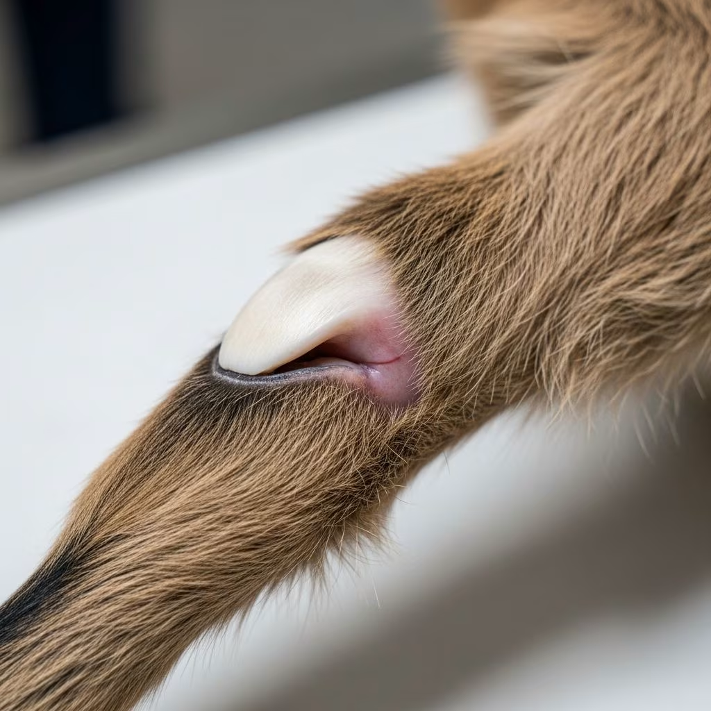
Epiphysitis represents a significant challenge in young goats, particularly those undergoing rapid growth phases, leading to disturbances in bone development at the growth plates. This condition often manifests in kids and first-time lactating does, resulting from nutritional excesses or deficiencies that disrupt calcium-phosphorus balance and other essential minerals.
Understanding the Pathophysiology of Epiphysitis
The growth plates, or physes, at the ends of long bones are critical for longitudinal bone growth in young goats. Epiphysitis occurs when these areas experience excessive pressure or nutritional stress, causing inflammation and potential permanent deformities. In goats, this is frequently tied to diets high in calcium relative to phosphorus, with ratios exceeding 1.4:1, often seen in alfalfa-heavy feeds.
Rapid growth exacerbates the issue, as seen in males more than females, and in young does under the strain of late pregnancy or heavy lactation. Indoor confinement limits sunlight exposure, reducing natural vitamin D synthesis, while overcast conditions compound this deficiency. Excess iron or protein in rations further impairs mineral absorption.
Recognizing Early Clinical Signs
Initial symptoms include subtle bowing of the radii, either laterally or medially, affecting one or both forelimbs. As the condition advances, goats display lateral deviation of the digits in fore or hind limbs, accompanied by lameness and reluctance to ambulate. An arched back becomes evident, signaling discomfort, alongside soft swellings and tenderness in key joints such as the carpus, metacarpophalangeal, tarsus, and metatarsophalangeal regions.
- Bowed forelimbs: Often the first visible change, progressing if untreated.
- Joint swelling: Soft and painful, distinct from infectious arthritis.
- Mobility issues: Goats may stand with an arched posture and avoid movement.
- Hoof abnormalities: Deviation in toe alignment on affected limbs.
These signs mimic other disorders like rickets or white muscle disease, necessitating precise diagnosis to avoid mis management.
Factors Contributing to Development
Several dietary and environmental elements converge to trigger epiphysitis. High-calcium feeds like alfalfa (1.4% calcium, 0.2% phosphorus) create imbalanced ratios, especially when kids are nursed on milk for extended periods due to limited market outlets.
| Risk Factor | Description | Impact on Goats |
|---|---|---|
| Calcium:Phosphorus Ratio >1.4:1 | Excess calcium from alfalfa or milk | Disrupts bone mineralization |
| High Protein Diets | Concentrates with unbalanced proteins | Accelerates growth unevenly |
| Vitamin D Deficiency | Indoor housing, poor feed stability | Impairs calcium absorption |
| Excess Iron | Contaminated feeds or water | Interferes with phosphorus uptake |
| Rapid Growth | Heavy milkers, twins/triplets | Overloads growth plates |
Carotene in feeds exerts an anti-vitamin D effect, and vitamin D instability in mineral-mixed rations worsens the scenario. Genetic selection for fast-maturing breeds may heighten susceptibility, akin to observations in rapidly growing foals.
Diagnostic Approaches for Confirmation
Veterinary assessment begins with a thorough history, focusing on diet, growth rate, housing, and reproductive status. Physical exams reveal characteristic swellings and pain on palpation. Radiography provides definitive confirmation, showing irregularities at the physes, distinguishing epiphysitis from osteochondrosis or infectious causes.
Blood analyses can quantify calcium, phosphorus, and vitamin D levels, while ruling out concurrent rickets. Differential diagnoses include caprine arthritis encephalitis (CAE), which causes chronic joint swelling but involves viral etiology and potential neurological signs.
Comprehensive Treatment Strategies
Correction hinges on addressing root causes. Dietary overhaul is paramount: reduce calcium-rich feeds, balance ratios to 1.2:1-2:1, and incorporate phosphorus supplements. Injectable vitamin D or oral balanced mineral mixes restore deficiencies.
- Nutritional Adjustment: Shift to growth-slowing rations with appropriate hay varieties, avoiding lush pastures.
- Supplementation: Phosphorus, vitamin D injections as prescribed.
- Management Changes: Promote exercise in sunlight; separate buck and doe kids by 3 months to prevent early breeding.
- Supportive Care: Anti-inflammatories for pain; monitor for self-correction in mild cases.
In young lactating does, enhanced nutrition supports bone remodeling, often reversing deformities over time. Prognosis improves with early intervention, halting progression.
Preventive Measures for Goat Herds
Proactive nutrition planning averts outbreaks. Formulate rations with verified Ca:P ratios, ensuring vitamin D fortification and stability. Discourage breeding does under 7-8 months, and provide ample sunlight exposure.
- Monitor growth rates in kids, intervening if excessive.
- Separate sexes early to avoid unplanned pregnancies.
- Regular deworming and mineral profiling via blood tests.
- Select breeding stock without epiphysitis history.
Herd-level strategies mirror those for developmental orthopedic diseases in other species, emphasizing balanced nutrition over rapid gains.
Long-Term Prognosis and Complications
Untreated epiphysitis leads to permanent limb angular deformities, compromising mobility and productivity. Early dietary corrections typically allow self-resolution, especially in growing kids. In does, deformities may persist but stabilize with proper support, preventing welfare issues.
Concurrent conditions like rickets amplify severity, underscoring holistic management. Regular radiographic follow-ups track progress.
FAQs on Epiphysitis in Goats
What is the most common cause of epiphysitis in goats?
Imbalanced calcium-to-phosphorus ratios in diets, often from high-alfalfa feeds or prolonged milk feeding.
Can epiphysitis be cured completely?
Yes, with prompt nutritional correction, most cases self-correct without lasting damage.
Is epiphysitis painful for goats?
It causes discomfort and lameness, but pain is moderate compared to infectious joint diseases.
How do you differentiate epiphysitis from CAE?
Epiphysitis shows growth plate changes on X-rays without viral signs; CAE is chronic and seropositive.
Should I supplement vitamin D routinely?
Only under veterinary guidance, as excess can harm; focus on balanced rations first.
Nutritional Guidelines Table for Prevention
| Nutrient | Recommended Level (Growing Kids) | Sources |
|---|---|---|
| Calcium | 0.6-1.0% | Balanced hay, minerals |
| Phosphorus | 0.4-0.6% | Grains, supplements |
| Ca:P Ratio | 1.2:1 to 2:1 | Formulated feeds |
| Vitamin D | 300-500 IU/kg | Sunlight, stabilized additives |
| Protein | 14-16% | Legume-grass mixes |
References
- How to treat epiphysitis — Farmer’s Weekly. 2019-12-05. https://www.farmersweekly.co.za/animals/horses/how-to-treat-epiphysitis/
- Epiphysitis in Goats — Merck Veterinary Manual. Accessed 2026. https://www.merckvetmanual.com/musculoskeletal-system/lameness-in-goats/epiphysitis-in-goats
- White Muscle Disease in Goats — MSD Veterinary Manual. Accessed 2026. https://www.msdvetmanual.com/en/Musculoskeletal-System/Lameness-in-Goats/White-Muscle-Disease-in-Goats
- Swollen Joints in Goats and Other Knee-Related Concerns — Goat Journal. Accessed 2026. https://goatjournal.iamcountryside.com/health/swollen-joints-in-goats/



