

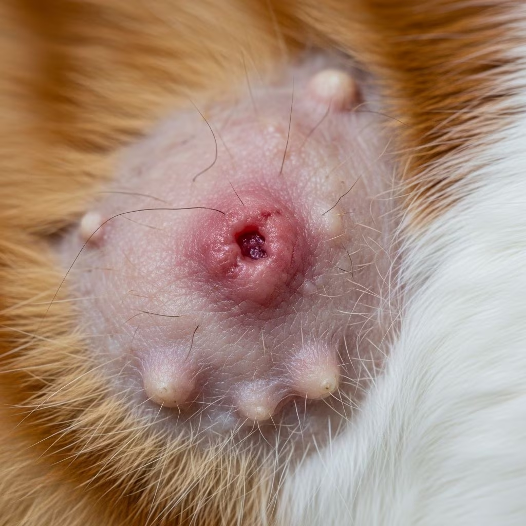
Feline eosinophilic skin disorders, commonly grouped under the umbrella of eosinophilic granuloma complex (EGC), represent a cluster of inflammatory conditions triggered primarily by hypersensitivity reactions. These disorders manifest as distinct skin lesions including plaques, granulomas, and indolent ulcers, often linked to allergies from fleas, food, or environmental factors. Understanding these conditions is crucial for cat owners to ensure prompt intervention and improved quality of life for their pets.
Understanding the Nature of Eosinophilic Conditions
Eosinophils are a type of white blood cell involved in combating parasitic infections and allergic responses. In cats, an overzealous accumulation of these cells in the skin leads to the characteristic lesions of EGC. While the precise etiology remains elusive, research points to an exaggerated immune response, potentially with genetic predispositions. These conditions are not contagious but can recur if underlying triggers persist, affecting cats of any age, though younger and middle-aged felines are more commonly impacted.
The term EGC encompasses three primary lesion types, each with unique presentations but sharing a common inflammatory pathway. Veterinary dermatologists emphasize that early recognition prevents progression and secondary complications like infections.
Key Manifestations of Feline Eosinophilic Skin Issues
Eosinophilic Plaques: Raised and Irritated Patches
These appear as well-defined, raised, red, and often moist areas on the skin, predominantly on the abdomen, inner thighs, or hind legs. Cats frequently groom excessively due to intense itching, leading to hair loss and potential ulceration. Plaques are hot to the touch and may ooze, signaling active inflammation driven by eosinophil infiltration.
Linear Granulomas: Elongated Swellings
Granulomas present as linear, ridge-like thickenings, commonly along the back legs, thighs, or even the tongue and palate. They feature hair loss, crusting, and a firm texture, sometimes resembling scars. Unlike plaques, they may be less pruritic but can cause discomfort if located in sensitive areas like the mouth.
Indolent Ulcers: Shallow Oral Lesions
Also known as rodent ulcers, these are shallow, erosive sores primarily on the upper lip, though they can extend to gums or nasal planum. They lack sharp borders, bleed easily, and resist healing without intervention. Despite the name, they are not always painful initially but can become so with progression.
| Lesion Type | Common Locations | Key Features | Itch Level |
|---|---|---|---|
| Plaques | Abdomen, thighs | Raised, red, moist | High |
| Granulomas | Hind legs, mouth | Linear, thickened | Moderate |
| Indolent Ulcers | Lips, gums | Shallow erosions | Low initially |
Triggers and Risk Factors Behind the Scenes
The most frequent instigators are flea allergy dermatitis, where even a single bite provokes a cascade of eosinophil recruitment. Food sensitivities to proteins like beef or fish, environmental allergens such as pollen or dust mites, and less commonly, mosquito bites also play roles. Genetic factors may heighten susceptibility, with some breeds like Burmese or Sphynx showing higher incidence. Stress can exacerbate flares, underscoring a multifactorial origin.
- Flea Hypersensitivity: Strict parasite control is foundational, as fleas amplify immune overreactions.
- Dietary Allergens: Novel protein diets often resolve cases linked to commercial foods.
- Environmental Culprits: Indoor allergens persist year-round, complicating management.
Diagnostic Approaches for Accurate Identification
Veterinarians begin with a comprehensive history, noting lesion onset, seasonal patterns, grooming habits, and flea exposure. Physical exams reveal lesion morphology, with cytology or fine-needle aspirates showing eosinophils. Skin biopsies provide definitive diagnosis, distinguishing EGC from mimics like fungal infections, neoplasia, or autoimmune diseases. Bloodwork rules out systemic involvement, while allergy testing or elimination diets pinpoint triggers.
Behavioral clues, such as nocturnal outdoor activity correlating with mosquito exposure, aid presumptive diagnosis. Advanced imaging or specialist referral ensures precision in refractory cases.
Comprehensive Treatment Strategies
Treatment targets inflammation control, trigger elimination, and symptom relief. Corticosteroids remain first-line, administered orally, topically, or via injections. Prednisolone at 1-2 mg/kg daily induces rapid remission, tapered to minimize side effects like weight gain or diabetes risk.
Cyclosporine offers a steroid-sparing alternative for chronic cases, modulating T-cell responses effectively. Supportive therapies include:
- Antibiotics for secondary bacterial infections.
- Antihistamines or essential fatty acids for adjunct itch relief.
- Hypoallergenic diets via 8-12 week trials.
- Flea preventives like selamectin or fipronil year-round.
Surgical excision is reserved for persistent, localized lesions unresponsive to medical therapy.
Long-Term Management and Prognosis
Most cats achieve excellent control with tailored plans, though recurrences necessitate lifelong vigilance. Regular vet check-ups monitor for flares, adjusting protocols as needed. Owner education on trigger avoidance enhances outcomes, with many cats enjoying normal lifespans. Severe or non-responsive cases benefit from dermatology specialists. Prognosis brightens with early detection and compliance.
Preventive Measures for Feline Skin Health
Proactive steps mitigate risks:
- Maintain rigorous flea control for all household pets.
- Introduce novel protein diets if allergies suspected.
- Minimize environmental allergens via frequent cleaning and air filtration.
- Monitor for early skin changes during grooming sessions.
Vaccination status and overall wellness support resilience against hypersensitivity.
Frequently Asked Questions (FAQs)
Can eosinophilic skin disorders be cured completely?
While not always curable, they are highly manageable with treatment, preventing severe episodes.
Is flea control alone sufficient?
No, comprehensive therapy addresses multiple potential triggers.
What if my cat licks steroid creams?
Use Elizabethan collars to prevent ingestion; systemic options may be preferable.
Are certain breeds more prone?
Yes, some like Burmese show predisposition, but any cat can be affected.
How long until improvement post-treatment?
Often within days for steroids, though full resolution takes weeks.
Word Count: 1678 (excluding HTML tags)
References
- Eosinophilic Granuloma Complex in Cats — VCA Animal Hospitals. Accessed 2026. https://vcahospitals.com/know-your-pet/feline-eosinophilic-granuloma-complex-in-cats
- Feline Eosinophilic Dermatitis — PDSA. 2020-08-01. https://www.pdsa.org.uk/pet-help-and-advice/pet-health-hub/conditions/feline-eosinophilic-dermatitis
- Eosinophilic Granuloma Complex — Cornell University College of Veterinary Medicine. Accessed 2026. https://www.vet.cornell.edu/departments-centers-and-institutes/cornell-feline-health-center/health-information/feline-health-topics/eosinophilic-granuloma-complex
- Eosinophilic Granuloma Complex in Cats — International Cat Care. Accessed 2026. https://icatcare.org/articles/eosinophilic-granuloma-complex-in-cats
- Eosinophilic Granuloma in Cats — Mission Veterinary Clinic. Accessed 2026. https://missionvet.com/eosinophilic-granuloma-in-cats/
- Feline Eosinophilic Dermatoses — PubMed (NCBI). 2014-04-01. https://pubmed.ncbi.nlm.nih.gov/24666659/
- Eosinophilic Granuloma Complex – Feline — Zoetis. Accessed 2026. https://www2.zoetis.co.uk/products-solutions/dermatology/eosinophilic-granuloma-complex



