

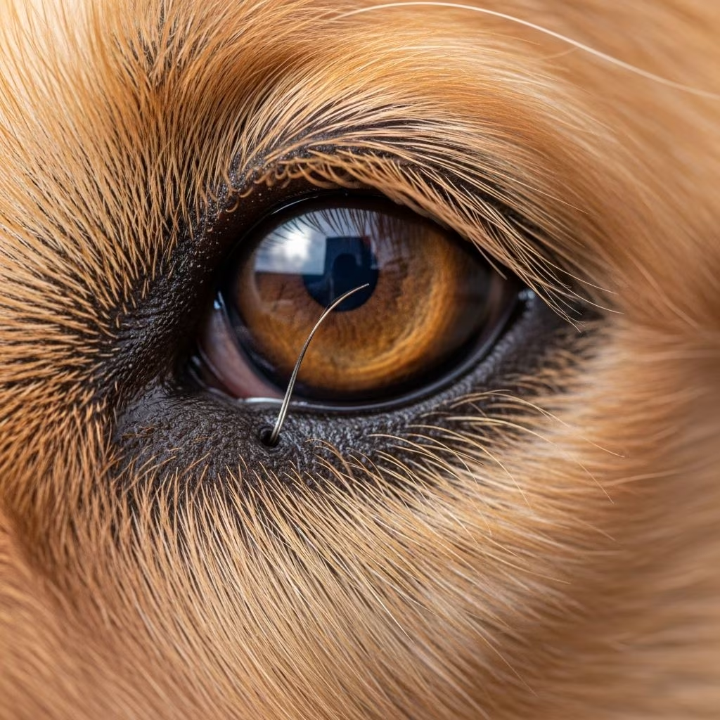
Understanding Ectopic Cilia in Dogs
Ectopic cilia represent a challenging eyelid condition that affects numerous dogs, particularly young animals. This inherited disorder involves abnormal hair growth that emerges through the conjunctiva, the delicate pink membrane lining the inside of the eyelid, rather than growing from the eyelid margin itself. These aberrant hairs grow inward and make direct contact with the cornea, the transparent front part of the eye, causing significant discomfort and potential complications if left untreated.
Unlike distichiasis, which involves extra eyelashes growing from the eyelid margin, ectopic cilia emerge directly through the conjunctival tissue. This anatomical difference makes ectopic cilia particularly problematic because the position of these hairs creates constant irritation against the sensitive corneal surface. The condition is genetic in nature, meaning affected dogs typically inherit the predisposition from their parents, and both eyes are commonly involved, though single-eye cases do occur.
Breed Susceptibility and Risk Factors
While ectopic cilia can affect any breed, certain dog breeds demonstrate increased susceptibility to this condition. Breeds more commonly affected include Bulldogs, Flat-coated Retrievers, Pekingese, English Cocker Spaniels, Rough Collies, Shetland Sheepdogs, and Yorkshire Terriers. The condition typically manifests in young dogs, particularly during their first few years of life, making early detection and intervention crucial for preventing serious ocular complications.
The genetic component of ectopic cilia means that breeding decisions should account for this condition’s presence in dog bloodlines. Responsible breeders often screen their breeding stock to minimize the transmission of this hereditary trait to subsequent generations, helping reduce the overall incidence of the disease in dog populations.
Recognizing Symptoms and Clinical Signs
Dogs suffering from ectopic cilia typically display several characteristic signs indicating ocular distress. Pet owners should remain vigilant for the following symptoms:
Common Clinical Presentations
– Excessive tearing or epiphora from the affected eye- Involuntary blinking or blepharospasm (frequent eye closing)- Redness and inflammation of the conjunctiva and sclera- Ocular discharge, ranging from clear to mucopurulent- Pawing, rubbing, or scratching at the face and eyes- Squinting or keeping the affected eye partially or fully closed- Visible corneal opacity or cloudiness- Corneal ulceration in advanced cases- Secondary signs including whining, depression, and behavioral changes- Bumping into objects due to vision impairment from keeping one eye closed- Reduced interest in play or outdoor activities
It is crucial to understand that ectopic cilia are almost always extremely painful for affected dogs. Unlike some cases of distichiasis where dogs may show no clinical signs, ectopic cilia virtually always cause discomfort due to their direct contact with the cornea. The severity of clinical signs may vary depending on the number of ectopic cilia present, their exact location, and individual corneal sensitivity.
Diagnostic Procedures and Examination
Accurate diagnosis of ectopic cilia requires a thorough veterinary eye examination, often necessitating sedation or general anesthesia for complete visualization. Veterinarians employ several diagnostic techniques to confirm the condition:
Diagnostic Methods
– **Direct observation:** Identifying abnormal lashes emerging from the eyelid or touching the cornea during physical examination- **Slit-lamp examination:** Using specialized magnification equipment to visualize small ectopic cilia that may not be visible to the naked eye- **Schirmer tear test:** Measuring tear production to assess the eye’s ability to maintain appropriate moisture levels- **Fluorescein dye test:** Applying orange dye to detect corneal abrasions and ulceration patterns- **General anesthesia examination:** Conducting comprehensive ocular evaluation under anesthesia when initial examination proves inconclusive
A characteristic diagnostic finding includes peripheral corneal ulcers in young dogs that fail to respond to conventional topical treatments. These non-healing or recurrent ulcers often signal the presence of ectopic cilia, particularly when observed in individuals predisposed to the condition. The ulcers typically appear as round lesions toward the edges of the cornea and may develop a distinctive appearance that helps differentiate them from other corneal pathologies.
Treatment Options for Ectopic Cilia
Treatment decisions for ectopic cilia depend on the severity of clinical signs, the number of abnormal hairs present, and the degree of corneal involvement. Several treatment approaches are available, ranging from conservative management to surgical intervention.
Non-Surgical Management
For dogs without clinical signs or those showing only mild symptoms, conservative treatment may be attempted initially:
– **Ocular lubricants:** Twice-daily application of lubricating gels or ointments helps improve tear film quality and may reduce irritation in mild cases. However, this approach requires lifelong commitment and provides temporary relief rather than permanent resolution.- **Topical antibiotics:** When secondary bacterial infections develop, antibiotic drops or ointments prevent further complications and promote corneal healing.- **Temporary plucking:** Using epilation forceps to remove offending hairs provides diagnostic confirmation and temporary relief, though hairs typically regrow every 4-6 weeks, often becoming thicker upon regrowth, making this unsuitable as a long-term solution.
Many veterinarians may attempt medical management in mild cases, but the temporary nature of these approaches means most dogs with ectopic cilia ultimately require surgical intervention for lasting comfort and prevention of vision-threatening complications.
Surgical Treatment Options
Surgical removal of ectopic cilia represents the most effective and recommended treatment approach, with multiple techniques available depending on individual case characteristics:
Cryoepilation (Cryotherapy)
Cryoepilation has emerged as the most widely recommended surgical technique for treating ectopic cilia. During this procedure, performed under general anesthesia, abnormal hair follicles are frozen using a liquid nitrogen probe, and the hairs are subsequently removed individually. This technique boasts an 85-90% success rate in most dog breeds when performed after three years of age. The procedure effectively destroys the hair follicle root, preventing regrowth in most cases. Shih Tzus and some other breeds show higher rates of hair regrowth post-operatively, sometimes necessitating repeat procedures.
Surgical Excision
Traditional surgical excision involves removing the abnormal hair along with its associated follicle under general anesthesia. The surgical site is left to heal by scarring, and provided the entire follicle is removed, permanent hair elimination typically occurs. This technique offers reliable results with minimal recurrence when performed correctly by experienced veterinarians.
Punch Biopsy Technique
A punch biopsy represents an alternative surgical technique particularly useful for removing ectopic cilia causing corneal ulcers. This method demonstrates good results in terms of ulcer healing and offers advantages in certain clinical scenarios.
Electrolysis
Electrolysis represents another surgical option that destroys hair follicles through electrical energy, though it may be less commonly employed than cryotherapy or traditional excision.
Surgical treatment should be pursued urgently when ectopic cilia cause significant corneal ulceration, as continued abrasion may lead to serious secondary complications requiring additional surgical corneal repair procedures. The decision regarding which surgical technique to employ should be made collaboratively between pet owners and veterinary specialists based on individual case factors.
Prognosis and Post-Operative Management
The prognosis for ectopic cilia following appropriate surgical treatment is generally excellent. Dogs experiencing no clinical signs enjoy an excellent long-term outlook, while those with mild signs managed medically typically respond well when treatment protocols are properly followed. Even for surgical cases, outcomes are favorable when the entire hair follicle is completely removed.
Post-operative care plays a vital role in ensuring successful recovery and preventing complications:
– **Antibiotic therapy:** Continue prescribed antibiotic drops or ointment as directed, applying drops before ointments with adequate interval between applications- **Eye cleansing:** Gently wipe away discharge using clean water and soft cotton wool before applying medications- **Activity restriction:** Limit strenuous activity during the healing period to prevent trauma to the surgical site- **Follow-up examinations:** Attend all scheduled post-operative rechecks for several months to monitor for potential hair regrowth- **Medication compliance:** Administer all prescribed medications consistently and for the complete recommended duration
While hair regrowth occurs in some cases, particularly in young dogs and certain breeds like Shih Tzus, the majority of dogs experience permanent resolution following appropriate surgical treatment. Associated corneal ulceration typically heals rapidly after surgery when no secondary infection has occurred.
Untreated Consequences and Long-Term Outcomes
Failing to address ectopic cilia can result in serious, potentially vision-threatening complications. Without treatment, severe corneal damage may develop, leading to corneal scarring, vision deterioration, and in severe cases, permanent blindness or loss of the affected eye. The progressive nature of untreated ectopic cilia means that early intervention significantly improves long-term outcomes and quality of life.
Frequently Asked Questions About Ectopic Cilia
Q: How are ectopic cilia different from distichiasis?
A: Ectopic cilia emerge through the conjunctiva on the inside of the eyelid, directly contacting the cornea, while distichiasis involves extra eyelashes growing from the eyelid margin. Ectopic cilia are almost always painful, whereas some distichiasis cases cause no clinical signs.
Q: Can ectopic cilia affect only one eye?
A: While both eyes are typically affected with this genetic condition, it is possible for only one eye to show clinical signs or be diagnosed initially. A thorough examination of both eyes is important during diagnosis.
Q: What is the success rate of surgical treatment?
A: Cryoepilation, the most recommended technique, demonstrates 85-90% effectiveness in most dog breeds when performed after age three. Success rates are excellent for complete surgical excision when the entire follicle is removed.
Q: Will my dog’s hair grow back after surgery?
A: In most cases, when the entire hair follicle is completely removed, permanent hair elimination occurs. However, some dogs, particularly young animals and certain breeds like Shih Tzus, may experience regrowth requiring repeat procedures.
Q: Is surgery necessary if my dog shows no symptoms?
A: Dogs without clinical signs may not require treatment initially. However, regular monitoring is essential, as symptoms may develop over time. Dogs with mild symptoms may be managed medically, while surgical intervention is typically recommended when significant discomfort or corneal ulceration occurs.
Q: How long is recovery after ectopic cilia surgery?
A: Most dogs recover well from surgery. Post-operative monitoring is necessary for several months to watch for potential hair regrowth. Follow-up veterinary examinations ensure proper healing and early detection of any complications.
References
- Distichiasis and Ectopic Cilia in Dogs — Embrace Pet Insurance. Accessed November 28, 2025. https://www.embracepetinsurance.com/health/distichiasis-and-ectopic-cilia
- Distichiasis or Ectopic Cilia (extra eyelashes) in dogs — Animal Trust. Accessed November 28, 2025. https://www.animaltrust.org.uk/pet-health-advice-hub/extra-eyelashes-in-dogs
- Ectopic Cilia — MVS Veterinary Services. Accessed November 28, 2025. https://www.mvsvets.com/wp-content/uploads/2015/08/Ectopic-Cilia.pdf
- ECTOPIC CILIA — Veterinary Vision. Accessed November 28, 2025. https://www.veterinaryvision.co.uk/images/factsheets/Veterinary-Vision-Factsheet-Ectopic-cilia.pdf
- Distichiasis or ‘extra eyelashes’ Fact Sheet — Veterinary Specialists. Accessed November 28, 2025. https://www.vetspecialists.co.uk/fact-sheets-post/distichiasis-extra-eyelashes-fact-sheet/
- Ectopic Cilia in Dogs – Symptoms, Causes, Diagnosis, Treatment — Wag Walking. Accessed November 28, 2025. https://wagwalking.com/condition/ectopic-cilia
- Ectopic cilia — EyeVet.ie. Accessed November 28, 2025. https://www.eyevet.ie/wp-content/uploads/2009/01/ectopic_cilia.pdf
- Ectopic cilia — Paragon Veterinary Referrals. Accessed November 28, 2025. https://www.paragonreferrals.co.uk/fact_sheets/ectopic-cilia/
- Eye ulcers in dogs caused by ectopic cilia. Excision by biopsy — Vets and Clinics. Accessed November 28, 2025. https://vetsandclinics.com/en/eye-ulcers-in-dogs-caused-by-ectopic-cilia-excision-by-biopsy



