

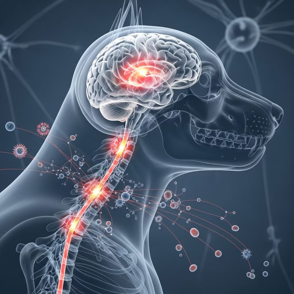
Inflammation affecting the brain and spinal cord represents one of the most serious neurological challenges veterinarians encounter in canine patients. When the protective membranes surrounding these vital structures become inflamed, dogs experience significant neurological dysfunction that can rapidly progress to life-threatening complications. Understanding the distinction between different types of neural inflammation, recognizing early warning signs, and knowing when to seek emergency veterinary care are essential for any dog owner. This guide provides detailed information about these conditions, their underlying causes, diagnostic approaches, and the therapeutic strategies available to help affected dogs.
Distinguishing Between Related Neurological Conditions
Several terms describe inflammatory conditions affecting the central nervous system, and understanding these distinctions is crucial for appropriate treatment. The membrane system surrounding the brain and spinal cord consists of protective layers called the meninges. When these membranes become inflamed, the condition is termed meningitis. When inflammation extends into the brain tissue itself, it is called encephalitis. When both structures are affected simultaneously, veterinarians use the term meningoencephalitis to describe the combined inflammation of both the protective membranes and the underlying neural tissue.
Each condition presents with overlapping but distinct clinical features. Meningitis primarily affects the protective layers, while encephalitis damages the functional neural tissue within the brain. The location and extent of inflammation significantly influence the specific symptoms a dog will develop, ranging from mild discomfort to severe neurological dysfunction.
Recognizing Clinical Manifestations in Affected Dogs
Dogs with inflammation of the meninges typically display a characteristic set of initial symptoms. The most common signs include elevated body temperature, neck pain that becomes apparent when the head is flexed downward, and muscle rigidity throughout the body. Dogs may exhibit painful muscle spasms or cramping sensations without showing obvious dysfunction of the brain or spinal cord. In the early stages of disease, some dogs may present with only these localized signs.
When inflammation extends to involve the brain tissue itself or becomes widespread throughout both structures, the clinical presentation becomes more complex and severe:
- Altered mental states ranging from depression and lethargy to agitation and behavioral changes
- Vision loss or complete blindness
- Weakness or partial paralysis affecting facial muscles or limbs
- Loss of balance and coordination with difficulty maintaining posture
- Repetitive circling or head tilting to one side
- Recurrent seizure activity or convulsions
- Difficulty swallowing or consuming food and water
- Progressive loss of consciousness potentially progressing to comatose states
- Incoordination and stumbling movements
- Nystagmus or involuntary eye movements
The severity and specific combination of symptoms depend on which brain regions are affected and the degree of inflammation present. Some dogs may present with one predominant symptom, while others display multiple neurological deficits simultaneously.
Underlying Causes of Neurological Inflammation
The etiology of meningitis and encephalitis in dogs falls into two primary categories: infectious and non-infectious inflammatory causes. Determining the underlying cause is essential because treatment approaches differ significantly between these categories.
Infectious Causes
Bacterial infections represent one category of infectious causes. When bacterial organisms invade the central nervous system, they trigger acute inflammatory responses. Various bacterial species can cause these infections, including Staphylococcus species and other pathogenic bacteria. The condition may result from systemic infections that spread to the nervous system or from localized infections such as ear infections or bone infections that extend into neural tissue.
Fungal infections constitute another significant infectious cause, particularly in certain geographic regions. Fungal organisms can spread through the bloodstream to establish infections within the central nervous system, causing chronic inflammation that may persist for extended periods.
Rickettsial organisms and protozoal parasites also cause neurological inflammation in dogs. Some protozoal infections are transmissible through tick vectors, making them more common in certain geographic areas. Viral infections, including rabies and canine distemper virus, can trigger severe inflammation of the meninges and brain tissue.
Non-Infectious Inflammatory Disorders
In many cases, particularly those termed idiopathic or of unknown origin, the inflammation results from the dog’s own immune system attacking neural tissue. These autoimmune conditions represent dysregulation of immune function where protective mechanisms become destructive. Granulomatous meningoencephalitis (GME) represents one recognized idiopathic inflammatory condition affecting multiple dog breeds. This condition occurs without identified infectious organisms and appears to involve abnormal immune response patterns.
Diagnostic Evaluation and Confirmation Procedures
Accurate diagnosis requires a systematic approach combining clinical evaluation with laboratory testing. Veterinarians typically begin with a thorough neurological examination to localize the inflammation within the nervous system. Blood work may reveal nonspecific changes such as fever, elevated white blood cell counts with increased immature neutrophil populations, indicating infection or inflammation.
The definitive diagnostic tool involves cerebrospinal fluid (CSF) analysis obtained through spinal tap or lumbar puncture. This procedure allows direct examination of the fluid surrounding the brain and spinal cord. CSF analysis reveals elevated protein levels, increased cell counts, and sometimes identifies causative organisms. Specific testing of CSF samples can detect antibodies against infectious agents, helping identify rickettsial, protozoal, or bacterial causes. Elevated immunoglobulin A (IgA) levels in the CSF may suggest specific inflammatory patterns associated with certain conditions.
Advanced imaging techniques including MRI (magnetic resonance imaging) can visualize inflammation within brain structures and help exclude other causes such as tumors or structural abnormalities. These imaging studies are particularly valuable in cases where encephalitis is suspected, as they can reveal the distribution and extent of brain involvement.
Treatment Approaches Based on Underlying Etiology
Managing Infectious Causes
When infections drive the inflammatory process, antimicrobial therapy forms the foundation of treatment. The specific antimicrobial agents selected depend on the identified or suspected organism:
- Bacterial infections require antibiotic therapy selected based on culture results when possible. Ampicillin represents a commonly recommended option, administered at specific doses to penetrate the blood-brain barrier and reach therapeutic concentrations within the central nervous system. Alternative antibiotics including chloramphenicol, metronidazole, and trimethoprim-sulfonamide can effectively penetrate neural tissue when systemic antibiotics fail.
- Fungal infections typically require prolonged antifungal medication therapy, often continuing for months or longer. Amphotericin B represents the primary antifungal agent used for CNS fungal infections, though the specific dosing regimen requires careful management to balance efficacy against potential toxicity.
- Protozoal and rickettsial infections respond to combination therapy with trimethoprim-sulfonamide and pyrimethamine administered over 4-8 weeks.
Treatment duration for infectious causes typically extends for minimum of several months, with some infections requiring lifelong management. The prolonged treatment period reflects the difficulty antimicrobial agents face in achieving adequate concentrations within the central nervous system and the chronic nature of some infections.
Addressing Immune-Mediated Inflammation
Non-infectious inflammatory conditions require fundamentally different treatment strategies focused on suppressing the aberrant immune response. High-dose corticosteroids, particularly prednisolone, form the cornerstone of therapy for immune-mediated conditions. Corticosteroids suppress immune function, reducing the inflammatory cascade driving neurological damage.
Initial therapy typically involves prednisolone administered at 4 mg/kg daily given by mouth or intravenously, with gradual dose reduction over subsequent months as clinical improvement occurs. This immunosuppressive approach often produces dramatic improvement in clinical signs as inflammation subsides. Treatment duration typically spans 3-6 months or longer, with some cases requiring extended therapy.
In cases of severe inflammation or recurrent disease, more potent immunosuppressive agents may be added to corticosteroid therapy. Medications such as azathioprine can be combined with corticosteroids, often given in alternating schedules to reduce individual drug doses while maintaining therapeutic benefit. These chemotherapy-type medications necessitate periodic blood testing to monitor for toxicity.
Radiation therapy has demonstrated promising results in certain cases, particularly those with focal involvement of specific brain regions. Treatment protocols involving divided dose schedules appear effective in controlling inflammatory progression in selected patients.
Supportive Care Measures
Regardless of the underlying cause, comprehensive supportive care enhances treatment efficacy: Pain management becomes essential, as meningitis causes significant neck pain and discomfort. Anticonvulsant medications control seizure activity when present. Intravenous fluid administration maintains hydration and nutritional status in dogs unable to eat or drink normally. Bed rest allows the body to direct energy toward recovery, as these conditions substantially deplete energy reserves over weeks of illness. Physical therapy may assist in recovery of motor function during the rehabilitation phase.
Prognostic Considerations and Long-Term Outcomes
The prognosis varies considerably based on multiple factors. Dogs with acute disease that receive prompt immunosuppressive treatment typically experience favorable outcomes, particularly when immune-mediated causes are identified and treated aggressively. However, untreated dogs tend to develop a chronic course with periodic relapses and remissions, eventually declining without intervention.
The location and extent of inflammation significantly influence outcomes. Dogs with focal involvement limited to specific brain regions, such as forebrain structures causing seizures, often have longer survival times exceeding 395 days. Conversely, dogs with disseminated inflammation throughout the nervous system have median survival times measured in weeks without treatment. Older dogs with certain biomarker patterns, particularly elevated CSF IgA levels with frequent relapses, tend to require prolonged therapy with less favorable long-term outcomes.
Permanent neurological damage can result from inflammation-induced destruction of neural tissue. The reversibility of clinical signs depends on whether inflammation caused destructive damage to nervous tissue or merely functional disruption. Early diagnosis and prompt treatment maximize the likelihood of complete recovery before irreversible damage occurs.
Monitoring Progress and Treatment Adjustments
Careful monitoring during treatment informs decisions about therapy continuation or modification. Cerebrospinal fluid cell counts serve as a sensitive indicator of treatment success, with normalization of these counts suggesting adequate inflammation control. Blood and CSF profiles should return toward normal values before discontinuing therapy. In dogs with immune-mediated disease, treatment is typically continued for a minimum of 6 months after clinical signs resolve and laboratory values normalize.
Treatment cessation in immune-mediated cases must occur gradually, as sudden discontinuation of corticosteroids invariably leads to rapid and dramatic clinical deterioration. Slow tapering allows the dog’s immune system to stabilize at lower levels of inflammatory response without triggering acute flare-ups.
When to Seek Emergency Veterinary Care
Dogs displaying signs suggestive of meningitis or encephalitis require urgent veterinary evaluation. Any combination of fever, neck pain, stiff movements, seizures, vision loss, or altered consciousness warrants immediate professional assessment. The rapid progression potential of these conditions makes early intervention critical for optimal outcomes.
References
- Meningitis and Encephalitis in Dogs–Treat for the Treatable — DVM360. 2024. https://www.dvm360.com/view/meningitis-and-encephalitis-dogs-treat-treatable-proceedings
- The Symptoms of Meningitis in Dogs — Veterinary Neurology & Imaging of the Chesapeake. 2024. https://www.vetneurochesapeake.com/vnioc-blog/the-symptoms-of-meningitis-in-dogs
- Meningitis and Encephalitis in Dogs — Merck Veterinary Manual. 2024. https://www.merckvetmanual.com/dog-owners/brain-spinal-cord-and-nerve-disorders-of-dogs/meningitis-and-encephalitis-in-dogs
- Meningitis and Encephalitis in Dogs — MSD Veterinary Manual. 2024. https://www.msdvetmanual.com/dog-owners/brain-spinal-cord-and-nerve-disorders-of-dogs/meningitis-and-encephalitis-in-dogs
- Meningitis and Encephalitis in Dogs and Cats — BluePearl Pet Hospital. 2024. https://bluepearlvet.com/medical-articles-for-pet-owners/meningitis-and-encephalitis-in-dogs-and-cats/
- Neurology: Encephalitis in Dogs & Cats — NC State College of Veterinary Medicine. 2024. https://hospital.cvm.ncsu.edu/services/small-animals/neurology/encephalitis/
- Meningoencephalitis of Unknown Cause — Fitzpatrick Referrals. 2024. https://www.fitzpatrickreferrals.co.uk/neurology/meningoencephalitis-of-unknown-aetiology-mua/



