

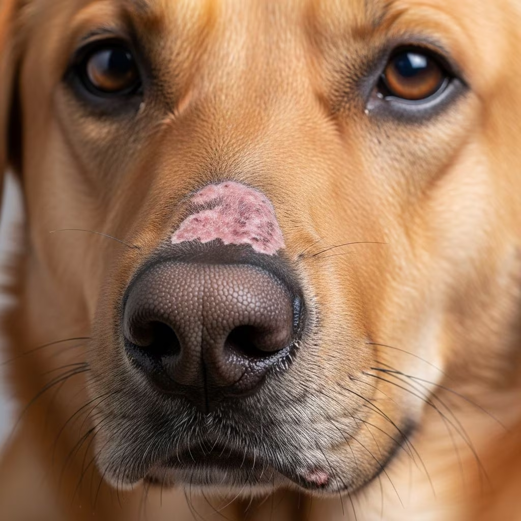
Discoid lupus erythematosus (DLE), often called cutaneous lupus, is a chronic autoimmune skin condition affecting dogs, leading to inflamed, scaly patches mainly on sun-exposed areas like the nose, ears, and face. This disease arises when the immune system mistakenly attacks healthy skin cells, causing discomfort, pigmentation changes, and potential scarring if unmanaged.
Understanding the Nature of Canine Discoid Lupus
DLE in dogs targets the skin without typically involving internal organs, distinguishing it from systemic lupus erythematosus (SLE). It manifests as well-defined, raised lesions with crusting and scaling, often progressing to depigmentation and ulceration. Early intervention is crucial to minimize permanent damage like nose leather loss or ear tip necrosis.
The condition’s autoimmune basis means genetic predispositions play a role, with certain breeds showing higher susceptibility. Environmental triggers exacerbate flares, making awareness vital for proactive care.
Breeds at Higher Risk and Genetic Factors
While DLE can affect any dog, it disproportionately impacts breeds such as German Shepherds, Shetland Sheepdogs, Border Collies, and Huskies. These herding and northern breeds may carry genetic markers heightening autoimmune responses.
- German Shepherds: Frequently reported due to thick coats contrasting with exposed skin vulnerabilities.
- Shetland Sheepdogs: Prone to facial lesions from light fur and sensitivity.
- Border Collies and Australian Shepherds: Active outdoor lifestyles increase UV exposure risks.
- Huskies and Malamutes: Pale skin under white fur amplifies sun damage.
Mixed breeds can also develop DLE, but purebred statistics highlight the need for breed-specific vigilance during routine check-ups.
Recognizing Early Symptoms of DLE
Initial signs often appear on the nasal planum (nose tip), presenting as redness, scaling, and crusting. Owners might notice depigmentation, giving the nose a chalky appearance, followed by cracking or ulceration.
| Stage | Symptoms | Common Locations |
|---|---|---|
| Early | Redness, mild scaling, itchiness | Nose, ears, eyelids |
| Progressive | Crusting, depigmentation, hair loss | Face, lips, paw pads |
| Advanced | Ulcers, scarring, secondary infections | Nasal planum, ear margins |
Other indicators include reluctance to eat hard kibble due to nasal pain, rubbing the face, or head shaking from ear discomfort. Unlike allergies, DLE lesions are sharply demarcated and worsen with sun exposure.
Causes and Triggers Behind DLE Development
The precise etiology remains multifactorial, combining genetic susceptibility with environmental insults. UV radiation is the primary trigger, prompting immune cells to attack keratinocytes in the basal epidermis.
Additional precipitants include:
- Prolonged sunlight without protection.
- Photosensitizing drugs or vaccines in rare cases.
- Stress, hormonal shifts, or concurrent infections weakening skin barriers.
Research indicates that in predisposed dogs, a single intense UV episode can initiate lesions, perpetuating a cycle of inflammation.
Veterinary Diagnosis: Confirming DLE
Diagnosis starts with a thorough history and dermatological exam. Vets assess lesion distribution and sun correlation. Cytology rules out infections, while a punch biopsy provides definitive histopathology: interface dermatitis, follicular atrophy, and basement membrane thickening.
Bloodwork screens for systemic involvement, checking ANA titers, though negative in pure DLE. Schirmer tear tests prevent keratoconjunctivitis sicca (KCS), a common comorbidity. Differential diagnoses include pemphigus, dermatomyositis, or fungal infections, necessitating expert evaluation.
Comprehensive Treatment Strategies
Treatment aims to suppress inflammation, promote healing, and avert scarring. No cure exists, but 80-90% of cases achieve remission with multimodal therapy.
Topical Therapies: First-Line Defense
High-potency corticosteroid ointments (e.g., fluocinolone) or tacrolimus apply directly to lesions, reducing erythema within 2-4 weeks. Intralesional injections offer targeted relief for thick plaques.
Systemic Medications for Moderate to Severe Cases
Oral options include:
- Tetracycline/niacinamide: Anti-inflammatory combo, minimizing steroid side effects.
- Hydroxychloroquine or cyclosporine: Immunomodulators for refractory disease.
- Short-term prednisone: For acute flares, tapered quickly.
Monitoring includes liver enzymes and blood counts.
Sun Protection Essentials
Daily SPF 30+ pet-safe sunscreen, UV-protective clothing, and shade during 10 AM-4 PM prevent flares. This non-pharmacologic pillar is mandatory.
Lifestyle Modifications for Long-Term Management
Beyond meds, holistic adjustments sustain remission:
- Regular vet visits every 3-6 months.
- Anti-inflammatory diet rich in omega-3s (fish oil).
- Avoid smoking environments (secondhand smoke worsens autoimmunity).
- Stress reduction via routine and enrichment.
These align with CDC lupus guidelines adaptable to canines.
Potential Complications and Prognosis
Untreated DLE risks squamous cell carcinoma on scarred noses (5-10% cases) and KCS leading to blindness. Most dogs enjoy good quality life with compliance; 20% may progress to SLE-like symptoms (joint pain, anemia). Prognosis excels with early detection—over 90% control achievable.
Preventive Measures for At-Risk Dogs
Proactive steps include genetic screening for breeders, lifelong sun avoidance, and prompt lesion reporting. Annual dermatology screens for predisposed breeds catch subclinical changes.
Frequently Asked Questions (FAQs)
Is discoid lupus in dogs contagious?
No, DLE is autoimmune, not transmissible to other pets or humans.
Can DLE resolve spontaneously?
Rarely; most require ongoing management to prevent relapse.
How long until treatments show results?
Topicals improve in 1-2 weeks; systemic in 4-6 weeks.
Does diet impact DLE?
Yes, omega-3 supplements reduce inflammation per studies.
Is surgery ever needed for DLE?
Only for severe scarring or cancer complications.
Recent Advances in Canine DLE Research
Emerging biologics like anti-TNF agents and JAK inhibitors show promise in trials for refractory cases. Photodynamic therapy aids non-responders. Ongoing studies refine genetic markers for early intervention.
References
- Discoid Lupus | Diagnosis & Disease Information — Rheumatology Advisor. 2023. https://www.rheumatologyadvisor.com/ddi/discoid-lupus/
- Discoid Lupus: Symptoms, Causes & Treatment Guide — Hightower Clinical. 2024. https://hightowerclinical.com/blogs/understanding-discoid-lupus/
- Understanding Discoid Lupus: Causes, Symptoms, and Treatments — Indiana University School of Medicine Dermatology. 2023. https://dermatrials.medicine.iu.edu/blogs/discoid-lupus-overview
- Discoid Lupus Erythematosus – StatPearls — NCBI Bookshelf. 2023-10-01. https://www.ncbi.nlm.nih.gov/books/NBK493145/
- Discoid Lupus Erythematosus: What Is It, Causes, Symptoms — Cleveland Clinic. 2024. https://my.clevelandclinic.org/health/diseases/21808-discoid-lupus



