

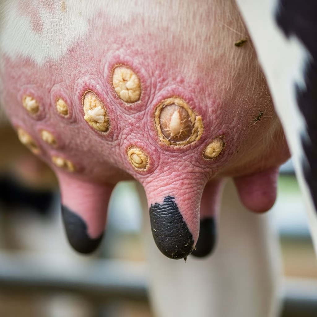
Cowpox represents a mild yet historically significant viral infection primarily targeting the skin of dairy cattle, with lesions concentrating on the udder and teats. Once widespread, it has become exceedingly uncommon, confined mostly to regions in western Europe. This poxvirus disease spreads through direct contact, especially during milking routines, underscoring the need for vigilant herd management.
The Viral Culprit Behind Cowpox
At its core, cowpox stems from a member of the orthopoxvirus genus, closely related to viruses causing smallpox and other pox infections. In cattle, it manifests as a self-limiting eruptive condition, where the virus invades epithelial tissues after entry through abrasions or mucous membranes. The pathogen thrives in dairy environments, exploiting the close physical interactions inherent to milking.
Historically, cowpox gained fame for conferring cross-immunity to smallpox in humans, a discovery pivotal to Edward Jenner’s vaccination breakthrough. Today, its rarity in cattle shifts focus to sporadic outbreaks, often linked to reservoir hosts like rodents. Genetic analysis reveals diverse strains, with cattle strains differing from those affecting cats or wildlife.
Recognizing Clinical Manifestations in Affected Animals
The disease unfolds after an incubation period of approximately 3 to 7 days, during which cattle may exhibit subtle fever. Initial signs include the emergence of papules on teats and udder skin, progressing rapidly to vesicles that often rupture, exposing raw ulcerated surfaces. These evolve into scabs, with full healing typically occurring within one month.
- Papular stage: Small, raised bumps appear first, signaling viral replication in skin layers.
- Vesicular and ulcerative phase: Fluid-filled blisters burst, leading to painful erosions prone to secondary bacterial invasion.
- Scabbing and resolution: Crusts form over defects, gradually sloughing as new epithelium regenerates.
In milking herds, infection rates can be high, affecting most animals due to shared milking equipment and handler contact. Affected cows rarely show systemic illness, maintaining milk production despite local discomfort. Differential diagnoses include pseudocowpox, bovine herpes mammillitis, and bacterial mastitis, necessitating lab confirmation.
Transmission Dynamics in Farm Settings
Cowpox propagates via mechanical transfer during milking, with virus-laden crusts and fluids contaminating hands, towels, or machines[10]. Calves and non-lactating cows are less commonly involved, as udder exposure predominates. Environmental persistence is notable; scabs retain infectivity for extended periods, resisting some disinfectants like alcohol but succumbing to bleach or heat above 80°C.
Zoonotic spillover occurs readily, with dairy workers developing similar lesions on hands, arms, or face following contact. Human cases feature fever, lymphadenopathy, and occasionally severe generalized illness, though fatalities are exceptional in immunocompetent individuals.
Diagnostic Approaches for Accurate Identification
Presumptive diagnosis relies on characteristic teat lesions in unvaccinated herds from endemic areas. Confirmation involves:
- Viral culture: From lesion swabs or scabs, grown in cell lines.
- PCR testing: Detects viral DNA with high sensitivity, ideal for non-viable samples.
- Histopathology: Reveals epidermal necrosis, eosinophilic inclusions, and dermal inflammation.
- Serology: Measures antibodies post-infection, useful for surveillance.
Electron microscopy visualizes brick-shaped poxvirus particles, aiding rapid preliminary assessment. Distinguishing cowpox from look-alike conditions prevents misdirected treatments.
Treatment Strategies and Supportive Care
Cowpox in cattle requires no specific antiviral therapy, as lesions resolve spontaneously. Management emphasizes:
| Aspect | Recommendations |
|---|---|
| Local care | Keep lesions clean; apply emollients to teats; isolate affected cows. |
| Secondary infections | Broad-spectrum antibiotics if bacterial complications arise. |
| Milking adjustments | Use separate utensils; milk last to minimize spread. |
| Human handlers | Gloves, handwashing; monitor for zoonotic signs. |
Healing timelines mirror cattle cases, spanning 4-6 weeks with scarring possible. Corticosteroids are strictly avoided, as they exacerbate viral spread in susceptible hosts.
Prevention and Control Measures for Herds
Vaccination against cowpox is obsolete given its scarcity, but biosecurity remains paramount. Key protocols include:
- Disinfect milking parlors with effective agents like quaternary ammonium compounds.
- Implement rodent control, as small mammals serve as reservoirs.
- Train staff on hygiene, promoting barrier protection.
- Surveillance via routine teat inspections during peak seasons.
Eradication efforts in Europe have marginalized the disease, but vigilance guards against re-emergence.
Zoonotic Implications and Public Health Concerns
Human cowpox, acquired from cattle or cats, presents as localized pox lesions with flu-like prodrome. High-risk groups include farmers and veterinarians. While self-limiting in healthy adults (resolving in 6-12 weeks), immunocompromised persons face graver outcomes.
No dedicated human vaccine exists post-smallpox eradication, but smallpox vaccine offers cross-protection. Prompt lesion isolation and contact precautions curb outbreaks.
Epidemiological Trends and Global Distribution
Confined to western Europe, cowpox in cattle reports dwindle annually. Spillover to cats now predominates, with rodents as amplifiers. Climate and wildlife dynamics may influence future patterns, warranting ongoing monitoring.
Comparative Pathology Across Species
While cattle experience mild udder-centric disease, felines suffer multifocal skin ulcers, potential pneumonia, and higher mortality if immunosuppressed. Cheetahs exhibit severe forms, highlighting host susceptibility variances.
FAQs on Cowpox in Cattle
What causes cowpox in dairy cows?
A poxvirus transmitted via milking contact, primarily affecting teats and udders.
How long does recovery take?
Lesions typically heal within 4 weeks without intervention.
Is cowpox contagious to humans?
Yes, handlers risk skin lesions and fever; use protective gear.
Can cowpox be prevented in herds?
Through hygiene, disinfection, and rodent management.
What’s the difference from pseudocowpox?
Cowpox lesions are larger, more ulcerative; lab tests differentiate.
Advanced Research Directions
Ongoing studies explore antivirals like tecovirimat for severe orthopox cases, though veterinary applications lag. Genomic sequencing tracks strain evolution, informing One Health strategies.
References
- Cowpox in Cattle — MSD Veterinary Manual. 2023. https://www.msdvetmanual.com/integumentary-system/pox-diseases/cowpox-in-cattle
- Cowpox Virus Infections in Cats and Other Species — Merck Veterinary Manual. 2023. https://www.merckvetmanual.com/integumentary-system/pox-diseases/cowpox-virus-infections-in-cats-and-other-species
- Cowpox — DermNet NZ. 2023. https://dermnetnz.org/topics/cowpox
- Cowpox Virus: Infectious Substances Pathogen Safety Data — Government of Canada, Public Health Agency. 2023. https://www.canada.ca/en/public-health/services/laboratory-biosafety-biosecurity/pathogen-safety-data-sheets-risk-assessment/cowpox-virus.html
- Cowpox Virus Infection in Cats — PMC (PubMed Central). 2023. https://pmc.ncbi.nlm.nih.gov/articles/PMC11148946/
- (Cow)pox virus — Langford Vets. 2022-01. https://www.langfordvets.co.uk/media/qp1dxtvm/poxvirus.pdf



