

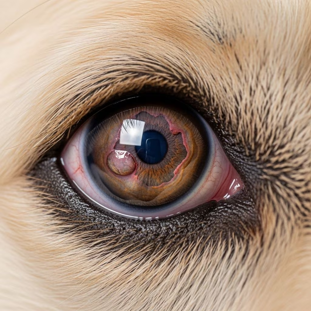
Corneal Ulcers in Dogs: A Comprehensive Guide
Corneal ulcers represent one of the most common eye conditions affecting dogs, requiring prompt recognition and appropriate treatment to prevent serious complications. The cornea, the transparent outer layer of the eye, plays a crucial role in vision and protection. When the corneal surface becomes damaged or eroded, it creates an ulcer that can range from superficial to deeply penetrating. Understanding this condition is essential for dog owners to recognize early signs and seek timely veterinary care.
What is a Corneal Ulcer?
A corneal ulcer is a loss of the corneal epithelium, which is the outermost protective layer of the eye. The cornea consists of several layers, and depending on which layers are affected, ulcers are classified into different categories. Superficial ulcers involve only the epithelial layer and typically heal quickly with appropriate treatment. More serious ulcers can extend into the stromal layer, the thicker middle portion of the cornea. A descemetocele represents the most severe type of corneal ulcer, where the ulceration extends nearly through the entire corneal depth, leaving only Descemet’s membrane—the innermost corneal layer—intact.
Causes of Corneal Ulcers in Dogs
Corneal ulcers develop through various mechanisms, each requiring specific management approaches. Traumatic injuries are among the most common causes, including scratches from vegetation, contact with foreign objects, or self-induced trauma from excessive eye rubbing. Dry eye syndrome, medically known as keratoconjunctivitis sicca, reduces the protective tear film and increases ulcer risk. Entropion, a condition where the eyelid rolls inward, causes constant eye irritation. Infections from bacteria, fungi, or viruses can directly cause ulceration or compromise the cornea’s integrity. Additionally, chemical burns, radiation exposure, and certain systemic diseases can contribute to ulcer formation. Understanding the underlying cause is critical for effective treatment and prevention of recurrence.
Clinical Signs and Symptoms
Dogs with corneal ulcers typically display obvious signs of ocular discomfort and irritation. The most common clinical manifestations include excessive tearing, eye redness, squinting or blepharospasm, and light sensitivity (photophobia). Affected dogs may hold their eye partially or completely closed and show reluctance to open it. Some dogs develop a cloudy or hazy appearance to the eye, particularly if the ulcer is deep or infected. Discharge from the eye may be present, ranging from clear and watery to purulent in cases of infection. In severe cases, dogs may exhibit behavioral changes such as increased anxiety, reluctance to eat, or general depression due to pain. Pet owners should seek immediate veterinary attention if they notice any combination of these signs, as prompt treatment significantly improves outcomes.
Diagnosis of Corneal Ulcers
Veterinarians employ several diagnostic techniques to identify corneal ulcers and determine their severity. The fluorescein dye test is the most common diagnostic tool, involving the application of specialized fluorescent dye drops to the eye. The dye adheres to areas where the corneal epithelium is absent, revealing the ulcer’s location and size under ultraviolet light. Additional diagnostic approaches may include cytology and culture if cellular infiltration or purulent discharge suggests infection. These tests help identify the specific microorganism involved, allowing for targeted antibiotic selection. Slit-lamp examination provides detailed visualization of the cornea’s depth and involvement of underlying layers. In some cases, confocal microscopy or other advanced imaging may be employed to assess stromal involvement. Careful examination is essential to distinguish between superficial ulcers and more serious conditions like descemetoceles, as these require dramatically different treatment approaches.
Treatment Options for Corneal Ulcers
Management of Superficial Corneal Ulcers
Corneal abrasions and superficial ulcers generally heal within three to five days with appropriate medical management. The primary treatment approach involves several key components working synergistically. Broad-spectrum topical antibiotics such as chloramphenicol serve as first-line therapy to prevent bacterial infections. These antibiotic drops or ointments must be applied regularly, typically 2-4 times daily or as directed by your veterinarian. Concurrent pain management is absolutely critical, as a painful eye prompts excessive rubbing and self-trauma that significantly delays healing. Oral non-steroidal anti-inflammatory drugs (NSAIDs) provide effective pain relief, while atropine sulfate or cyclopentolate hydrochloride eye drops serve dual purposes by relaxing the iris muscle and reducing reflex uveitis associated with corneal injury. Lubricating agents containing hyaluronate or similar substances maintain corneal moisture and promote healing throughout the recovery period.
One essential component of superficial ulcer management is the Elizabethan collar (E-collar or cone), which prevents the dog from rubbing or scratching the affected eye during the vulnerable healing phase. Even brief moments of self-trauma can significantly complicate healing and convert a simple ulcer into a more serious condition. Dogs should be kept away from bright light when receiving medications that dilate the pupil, as this causes discomfort and increased light sensitivity. Owners should monitor their dogs closely for signs of deterioration and attend follow-up appointments at five to seven days post-diagnosis to ensure appropriate healing progress.
Advanced Medical Therapy for Complex Ulcers
More complicated corneal ulcers that show slow healing or signs of infection require intensified medical management. Treatment protocols may include more potent topical antibiotics applied at higher frequencies, potentially every 2-4 hours depending on the specific diagnosis. Systemic oral antibiotics often complement topical applications in these cases. Serum therapy represents an innovative treatment approach for ulcers showing slow healing progression. Serum derived from blood contains valuable healing nutrients and proteins that inhibit stroma breakdown and promote tissue repair. Application of topical mucolytics such as N-acetylcysteine may be necessary if severe discharge accumulates on the corneal surface.
Grid keratectomy, a minor surgical procedure involving removal of loose epithelium that is not adhering properly, may be performed to freshen ulcer edges and encourage healing. This debridement process removes dead or poorly healing tissue layers that impede the normal healing response. Contact lenses are sometimes fitted to protect the outer eye layer during the healing process, particularly for uncomplicated ulcers. Dogs with uncomplicated ulcers receiving appropriate medical therapy typically heal within 3-7 days, though careful monitoring remains essential to prevent progression to more serious conditions.
Managing Pain and Discomfort
Effective pain management is fundamental to successful corneal ulcer treatment and cannot be overlooked. Systemic NSAIDs effectively reduce pain and inflammation, while additional medications such as paracetamol or opioid-based drugs like buprenorphine provide supplementary analgesia when necessary. Sedatives or anxiolytics including tramadol, acepromazine, or gabapentin may be prescribed if the dog is likely to cause further trauma through excessive activity or eye rubbing. Topical anesthetics are sometimes used temporarily to numb the cornea during diagnostic procedures, but these drugs are toxic to corneal tissue and can delay healing, so they should only be used once with caution. Atropine eye drops provide pain relief by dilating the pupil and relaxing intraocular muscles while reducing reflex uveitis.
Surgical Treatment for Severe Corneal Ulcers
Patients with perforation, descemetoceles, or deep ulcers extending approximately half the corneal depth or greater require urgent surgical intervention to prevent permanent blindness or eye loss. These serious cases necessitate prompt referral to a veterinary ophthalmologist who can determine whether the eye is salvageable and select the most appropriate surgical approach. Multiple surgical techniques exist for addressing severe corneal ulcers, each suited to specific clinical situations.
Corneo-limbal conjunctival transposition involves mobilizing tissue from the junction between the cornea and sclera along with conjunctival tissue to cover the ulcerated area, providing protection and supporting tissue healing. Corneal grafts replace severely damaged corneal tissue with healthy donor material, restoring corneal integrity and optical function. Conjunctival flaps, including both free grafts and pedicled flaps attached to blood vessels, are harvested from surrounding conjunctival tissue and sutured over the ulcerated area. Third eyelid flaps utilize the nictitating membrane to protect the injured cornea during healing. These surgical approaches protect the underlying defect, reduce pain, and provide an optimized environment for tissue regeneration. Regular monitoring during recovery is essential to ensure proper graft integration and to identify any complications requiring adjustment of the treatment protocol.
Managing Post-Ulcer Complications
After corneal ulcers heal, certain complications may develop that require additional management. Neovascularization—the growth of new blood vessels into the cornea—typically accelerates ulcer healing and is generally beneficial during the acute phase. However, once the ulcer has completely healed, extensive neovascularization can obstruct vision if not minimized. Your veterinarian may prescribe topical corticosteroid drops or ointments for several days to several weeks following complete ulcer healing to reduce the prominence and extent of these new vessels. This treatment helps preserve optimal vision while still allowing sufficient vascularization to support corneal health.
Monitoring and Follow-Up Care
Close monitoring during and after corneal ulcer treatment is absolutely essential to ensure appropriate healing and prevent serious complications. Owners should watch carefully for increased ocular discomfort, changes in discharge character or amount, or development of white, yellow, grey, or green infiltrates in the cornea, which could indicate deeper stromal infection. Any of these warning signs demand prompt re-examination and potential adjustment of the treatment plan. Fluorescein staining may be repeated at follow-up visits to monitor ulcer healing progress and identify any deterioration indicating the ulcer is becoming larger or deeper rather than resolving. If an ulcer isn’t healing as expected despite appropriate treatment, your veterinarian may change medications, perform swab cultures to identify specific microorganisms, or debride the ulcer edges to encourage healing.
Prevention of Corneal Ulcers
While not all corneal ulcers are preventable, several measures can significantly reduce risk. Regular eye examinations help identify predisposing conditions like dry eye syndrome or entropion before ulcers develop. Dogs with known dry eye should receive ongoing tear supplementation to maintain the protective tear film. Dogs prone to eye trauma should be supervised during outdoor activities and protected from vegetation or sharp objects. For dogs with entropion or other lid abnormalities, surgical correction can prevent chronic irritation. Prompt treatment of eye infections and careful wound care following ocular trauma reduce ulcer development risk. Elizabethan collars should be used immediately following any eye injury to prevent self-trauma during the critical initial healing phase.
Frequently Asked Questions
Q: How long does it take for a corneal ulcer to heal?
A: Superficial corneal ulcers and abrasions typically heal within 3-7 days with appropriate medical treatment. More serious ulcers involving deeper stromal layers may require 2-4 weeks or longer, and some may require surgical intervention for optimal outcomes.
Q: Can corneal ulcers lead to blindness?
A: Yes, if left untreated or inadequately managed, severe corneal ulcers can result in permanent scarring, perforation, or eye loss. Prompt veterinary treatment is critical to prevent these serious complications.
Q: Should I use over-the-counter eye drops for my dog’s corneal ulcer?
A: No. Over-the-counter eye drops may contain ingredients that harm corneal healing or worsen the condition. Only use medications prescribed by your veterinarian, as inappropriate treatment can significantly complicate recovery.
Q: Why does my dog need to wear a cone during corneal ulcer treatment?
A: The Elizabethan collar prevents your dog from rubbing or scratching the affected eye, which would cause additional trauma and significantly delay healing or convert a simple ulcer into a more serious condition.
Q: Can corneal ulcers recur?
A: Yes, particularly if the underlying cause isn’t addressed. For example, untreated dry eye or entropion can lead to recurrent ulcers. Your veterinarian will recommend preventive measures based on the cause of your dog’s initial ulcer.
Q: What is the difference between a corneal abrasion and a corneal ulcer?
A: A corneal abrasion is a superficial loss of the outer epithelial layer caused by trauma, while a corneal ulcer involves a deeper defect that may penetrate multiple corneal layers. Both conditions require treatment, but ulcers may need more intensive management.
References
- Corneal ulcers in dogs — Vet Times. 2024. https://www.vettimes.com/news/vets/small-animal-vets/corneal-ulcers-in-dogs
- Dog Corneal Ulcer: Symptoms and Treatment — Vets4Pets. 2024. https://www.vets4pets.com/pet-health-advice/dog-advice/corneal-ulcers-in-dogs/
- Corneal Ulcers in Dogs — VCA Animal Hospitals. 2024. https://vcahospitals.com/know-your-pet/corneal-ulcers-in-dogs
- Corneal Ulcers in Dogs: Medication vs. Surgery — Port City Veterinary Referral Hospital. 2024. https://www.portcityvet.com/blog/corneal-ulcers-in-dogs
- Eye ulcers (corneal ulcers) in dogs — PDSA. 2024. https://www.pdsa.org.uk/pet-help-and-advice/pet-health-hub/conditions/eye-ulcers-corneal-ulcers-in-dogs
- Eye Ulcer in Dogs: Medical Therapy — PetMD. 2024. https://www.petmd.com/dog/conditions/eyes/c_dg_Keratitis_Ulcerative
- Disorders of the Cornea in Dogs — MSD Veterinary Manual. 2024. https://www.msdvetmanual.com/dog-owners/eye-disorders-of-dogs/disorders-of-the-cornea-in-dogs



