

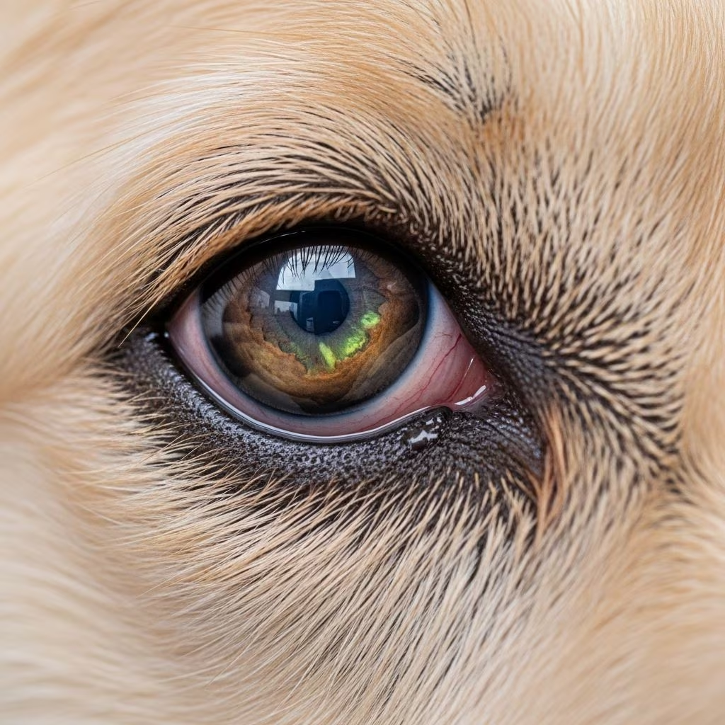
The cornea serves as a protective barrier for your dog’s eyes, but this delicate structure is susceptible to injury and disease. When the corneal surface becomes damaged, a painful condition known as a corneal ulcer develops. Understanding this condition is essential for any dog owner, as early recognition and prompt treatment can prevent serious complications and preserve your pet’s vision.
What Constitutes a Corneal Ulcer
A corneal ulcer is fundamentally a wound or abrasion that develops on the surface of the cornea, the transparent dome-shaped structure covering the front of the eye. This condition is medically referred to as ulcerative keratitis and causes inflammation and swelling of the corneal tissue. The cornea itself is composed of three distinct layers, each playing a critical role in maintaining eye function.
The outer layer, known as the epithelium, serves as the eye’s first line of defense against environmental hazards. Beneath this lies the stroma, a thicker supportive layer that maintains the cornea’s structural integrity and refractive properties. The deepest layer, Descemet’s membrane, works to contain the aqueous humor that bathes the interior structures of the eye.
Understanding these layers is important because the depth of an ulcer determines its severity and treatment requirements. Superficial ulcers that affect only the epithelium typically respond well to conservative treatment, while deeper ulcers that penetrate into the stroma demand more aggressive intervention.
Primary Causes of Corneal Ulceration in Canines
Corneal ulcers develop through various mechanisms, with trauma being the predominant culprit. However, understanding the full spectrum of causes allows owners to take preventative measures and recognize risk factors in their own dogs.
Traumatic Injury Mechanisms
Physical trauma represents the leading cause of corneal ulcers in dogs. This encompasses a wide range of scenarios that dog owners should recognize:
- Contact with sharp objects such as sticks, thorns, or grasses encountered during outdoor activities
- Scratch injuries from feline encounters or inter-dog conflicts
- Self-inflicted abrasions from the dog rubbing its eye excessively on carpets or furniture
- Blunt force trauma from running into objects or motor vehicle accidents
- Chemical irritation from substances like shampoo, drywall dust, or cleaning products entering the eye
Underlying Health Conditions
Beyond direct injury, several systemic and ocular health conditions predispose dogs to corneal ulceration:
- Keratoconjunctivitis Sicca (Dry Eye): Reduced tear production leaves the cornea vulnerable to damage and impairs healing mechanisms
- Eyelid Abnormalities: Conditions like entropion (inward-rolling eyelid) and distichiasis (abnormal eyelashes) cause chronic irritation
- Epithelial Dystrophy: An inherited corneal weakness particularly common in boxer breeds
- Endocrine Disorders: Cushing’s disease, diabetes, and thyroid dysfunction can compromise ocular health
- Facial Nerve Paralysis: Impaired eyelid movement prevents adequate eye protection
- Brachycephalic Anatomy: Flat-faced breeds like pugs and bulldogs face increased ulcer risk due to eye protrusion
Idiopathic Ulceration
Some dogs develop corneal ulcers without an identifiable cause, a condition termed indolent ulceration or spontaneous chronic corneal epithelial defects (SCCED). This condition most frequently affects geriatric dogs older than six years.
Recognizing Symptoms in Your Dog
Corneal ulcers are exceptionally painful, even when superficial. Dogs experiencing this condition often display behavioral and physical changes that warrant immediate veterinary evaluation.
Early Warning Signs
In the initial stages, observation may reveal:
- Excessive blinking or persistent eye closure
- Increased tear production or ocular discharge
- Redness and swelling around the eye
- Sensitivity to light
- General irritability or discomfort
Advanced Symptomatology
As ulcers progress or become more severe, additional indicators emerge:
- Visible cloudiness or blue-white discoloration of the cornea
- Yellow or green purulent discharge suggesting bacterial infection
- Behavioral changes such as hiding or appetite loss
- Visible depression or indentation on the eye surface
- Bulging of the cornea indicating structural weakness
Any of these severe symptoms constitute a veterinary emergency and require same-day professional evaluation.
Diagnostic Approaches
Veterinary diagnosis of corneal ulcers involves specialized examination techniques. Ophthalmologists typically apply fluorescein dye to the eye surface; damaged epithelium will retain the stain, creating a visible mark that clearly delineates the ulcer’s extent and severity. Additional imaging and examination methods help determine the underlying cause and assess whether deeper corneal layers are involved.
Treatment Strategies for Different Ulcer Severities
Treatment approaches vary significantly based on ulcer depth and complexity. A graduated response ensures appropriate care intensity matched to the condition’s severity.
Managing Superficial Ulcers
Superficial ulcers involving only the epithelial layer typically resolve within three to ten days with conservative management. Standard treatment includes:
- Topical antibiotic ointments or drops applied multiple times daily to prevent secondary bacterial infection
- Oral pain medications to minimize discomfort and improve quality of life
- Elizabethan collar (cone) to prevent self-trauma during healing
- Activity restriction to minimize eye irritation from environmental exposure
Intermediate Ulcer Management
Ulcers that show slow healing or moderate depth require more intensive pharmaceutical intervention:
- Frequent topical antibiotic application to prevent infection and promote healing
- Oral antibiotics when bacterial involvement is suspected or confirmed
- Anti-inflammatory medications to reduce corneal edema
- Corneal protective agents to support the healing process
- Possible minor surgical procedures such as keratotomy to stimulate healing
Severe Ulcer and Advanced Interventions
Deep ulcers or those approaching full corneal thickness demand aggressive management to prevent catastrophic outcomes:
- Intensive topical antibiotic regimens administered multiple times throughout the day
- Systemic antibiotics via oral or injectable routes
- Corneal protective medications that slow tissue deterioration
- Autologous serum drops created from the dog’s own blood to facilitate healing and prevent stromal collapse
- Surgical intervention including corneal grafting to replace damaged tissue and prevent rupture
Specialized Approaches for Dry Eye-Related Ulcers
When corneal ulcers result from keratoconjunctivitis sicca, treatment focuses on addressing the underlying tear deficiency. The primary therapeutic approach involves topical lacrimal stimulants such as tacrolimus or cyclosporine applied to the eye surface. These medications serve dual purposes—stimulating natural tear production while simultaneously providing lubrication. For severe dry eye cases, 0.03% tacrolimus in ointment form applied three times daily represents an effective standard regimen.
Post-Treatment Care and Prevention
Recovery from corneal ulcers requires diligent home care and monitoring. Dogs must wear protective Elizabethan collars throughout the healing period to prevent rubbing or scratching. Activity should be restricted to minimize exposure to irritants and reduce eye strain. Owners should administer medications precisely as prescribed and watch for signs of deterioration such as increased discharge, persistent cloudiness, or behavioral changes indicating worsening pain.
Preventing future ulcers involves identifying and addressing underlying predisposing factors. Dogs with dry eye require long-term management with tear stimulants. Those with eyelid abnormalities may benefit from surgical correction. Owners should minimize exposure to environmental hazards and promptly treat any ocular irritation before it progresses to ulceration.
Common Questions About Canine Corneal Ulcers
How quickly can a corneal ulcer develop?
Corneal ulcers can develop within hours of injury. A single traumatic event such as a cat scratch or thorn contact can create a full ulcer within a very short timeframe.
Can corneal ulcers heal without veterinary treatment?
Very superficial ulcers might show some improvement naturally, but professional evaluation and treatment dramatically improve healing speed and prevent serious complications including infection and permanent scarring.
Will my dog regain normal vision after an ulcer heals?
Vision outcomes depend on ulcer severity and depth. Superficial ulcers typically heal without permanent visual effects, while deep ulcers or those involving the stroma may result in scarring and reduced vision.
Are certain dog breeds more susceptible?
Brachycephalic breeds with prominent eyes face increased risk, as do breeds predisposed to epithelial dystrophy such as boxers. Older dogs are more prone to idiopathic ulceration.
References
- Corneal Ulceration in Dogs and Cats: Diagnosis and Treatment — VetBloom. 2024. https://vetbloom.com/corneal-ulceration-in-dogs-and-cats/
- Corneal Ulcers in Dogs — Animal Eye Surgery Center. 2024. https://www.animaleyeguys.com/site/blog/2024/07/30/corneal-ulcers-dogs
- Dog Eye Ulcer: Causes, Symptoms and Treatment Options — CareCredit. https://www.carecredit.com/well-u/pet-care/corneal-ulcer-in-dogs/
- Corneal Ulcers in Dogs — VCA Animal Hospitals. https://vcahospitals.com/know-your-pet/corneal-ulcers-in-dogs
- Corneal Ulcers in Dogs: Symptoms, Treatment & More — Hill’s Pet Nutrition. https://www.hillspet.com/dog-care/healthcare/corneal-ulcers-in-dogs
- Corneal Ulcers in Dogs — Premier Veterinary Group. https://www.premiervets.net/blog/corneal-ulcers-in-dogs



