

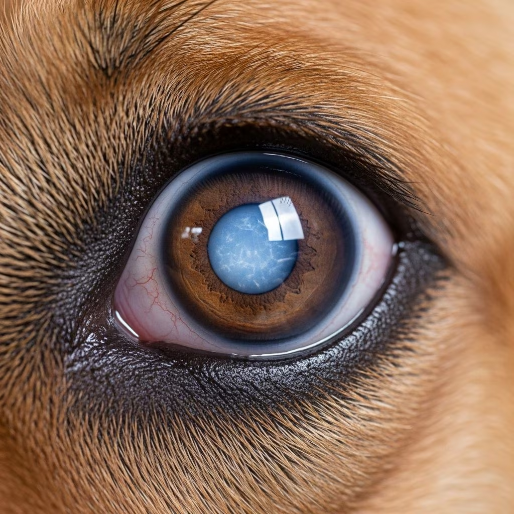
Understanding Corneal Lipidosis in Dogs
Corneal lipidosis is an accumulation of fatty substances, typically cholesterol, within the various layers of the cornea. The cornea is the transparent outer layer of the eye that allows light to enter and helps focus vision. When lipid deposits accumulate in this normally clear tissue, they create an opacity that can affect the appearance of your dog’s eye. Understanding this condition is essential for dog owners, particularly those with breeds predisposed to developing corneal lipidosis.
This eye condition can range from asymptomatic and benign to progressive and visually significant, depending on its underlying cause and severity. While some dogs experience no symptoms or vision loss, others may develop complications requiring veterinary intervention. Early recognition and proper management are crucial for maintaining your dog’s eye health and quality of life.
What Causes Corneal Lipidosis?
Corneal lipidosis develops through three distinct mechanisms, each with different implications for your dog’s health and treatment approach.
Corneal Dystrophy
Corneal dystrophy represents an inherited genetic condition where lipid deposits form within the corneal layers. This hereditary form is most commonly observed in dogs and rarely affects cats. The condition typically presents bilaterally, meaning it affects both eyes equally. Dogs with corneal dystrophy due to genetic predisposition experience painless lipid accumulation with minimal effects on vision. Several breeds show particular susceptibility to this inherited condition, including beagles, Cavalier King Charles spaniels, Siberian huskies, Alaskan malamutes, Samoyeds, American cocker spaniels, Labrador retrievers, and collies. The lipid typically consists of cholesterol, neutral fats, and phospholipids deposited in the subepithelial or stromal layers of the cornea.
Corneal Degeneration
Unlike the inherited form, corneal degeneration occurs as a secondary development following inflammation or injury to the eye. This type of lipidosis develops in response to other underlying eye diseases and inflammatory conditions. Anterior uveitis, keratitis, keratoconjunctivitis sicca (commonly known as “dry eye”), and scleritis can all trigger lipid accumulation as a secondary response. Additionally, corneal trauma such as ulceration that healed with lipid deposits can lead to corneal degeneration. This form appears more frequently in dogs than in cats and requires identification and treatment of the primary eye condition.
Elevated Blood Cholesterol Levels
Systemic hyperlipidemia and metabolic diseases can contribute to corneal lipidosis development. Hypothyroidism represents a particularly significant metabolic condition associated with lipid accumulation in the cornea. When dogs experience elevated cholesterol or triglyceride levels in their bloodstream, lipid can infiltrate corneal tissues, creating deposits that may progress over time. This type of lipidosis underscores the importance of screening for systemic diseases when corneal lipidosis is diagnosed.
Recognizing Symptoms and Clinical Signs
The clinical presentation of corneal lipidosis varies considerably depending on the underlying cause and severity of the condition.
Inherited Corneal Dystrophy Presentation
Dogs with inherited corneal dystrophy typically display whitish or grayish deposits with a granular or ground glass appearance within the cornea. These deposits typically appear in the central or paracentral region of the cornea and usually present as circular or oval lesions. Most importantly, inherited corneal dystrophy does not cause pain or vascularization (development of new blood vessels) in the affected cornea. Many dogs with this condition show no obvious external signs of discomfort and maintain normal vision.
Secondary Lipid Infiltration Signs
When lipidosis develops secondary to inflammation, trauma, or systemic disease, the clinical presentation differs significantly. White deposits appear on the eye following corneal injury, and these lesions typically produce irritation and become vascularized with new blood vessel development. Secondary lipid infiltration can result from conditions such as limbal melanoma, episcleritis, pannus, or abnormal new blood vessel formation that leaks lipid into corneal tissue. Unlike the painless nature of inherited dystrophy, secondary lipidosis often causes discomfort due to accompanying inflammation.
Diagnosis of Corneal Lipidosis
Accurate diagnosis requires a comprehensive approach combining clinical examination, laboratory investigation, and patient history evaluation.
Thorough Eye Examination
Your veterinarian will perform a detailed ophthalmic examination to assess the cornea and surrounding structures. This examination typically includes fluorescein dye application, which helps identify corneal ulcerations or epithelial damage that might accompany lipid deposits. The Schirmer tear test measures tear production and helps rule out keratoconjunctivitis sicca as a contributing factor. Careful assessment determines the location, extent, and characteristics of lipid deposits.
Patient History and Breed Considerations
Your veterinarian will gather detailed information about your dog’s age, breed, and medical history. Breed predisposition helps determine whether inherited dystrophy is likely. Age of onset provides valuable diagnostic information, as hereditary forms typically appear at specific developmental stages. A history of previous eye trauma, surgery, or systemic illness guides investigation toward secondary causes.
Laboratory Testing
When your veterinarian suspects hyperlipidemia or metabolic disease, blood work becomes essential. Fasting cholesterol and triglyceride levels reveal systemic lipid abnormalities. Blood glucose testing helps identify diabetes as a potential contributing factor. Thyroid function testing screens for hypothyroidism, which frequently correlates with corneal lipidosis. These laboratory results guide treatment decisions and help prevent progression of corneal disease.
Treatment Approaches for Corneal Lipidosis
Treatment strategies vary substantially based on the underlying cause and severity of corneal lipidosis.
Management of Inherited Corneal Dystrophy
Dogs with inherited corneal dystrophy typically require no active treatment since the condition rarely causes pain or significant vision impairment. Instead, your veterinarian will recommend periodic monitoring through routine eye examinations to watch for potential complications such as corneal ulcerations. This conservative approach reflects the generally benign nature of hereditary lipidosis in most affected dogs. Your veterinarian will establish an appropriate monitoring schedule based on your individual dog’s condition.
Treatment of Secondary Corneal Degeneration
When lipidosis develops secondary to inflammation or other eye diseases, treatment focuses on managing the primary condition. Antibiotics or anti-inflammatory eye drops address underlying infection or inflammation. Pain medications provide comfort if the corneal surface becomes irregular or ulcerated. Artificial tear ointments offer lubrication and protect the corneal surface from further damage. In some cases, additional therapeutic interventions may be necessary depending on the specific underlying condition.
Managing Metabolic Contributors
For dogs with corneal lipidosis related to systemic hyperlipidemia, dietary modifications play an important role. High-fat diets, treats, and nutritional supplements should be avoided or minimized. Treatment of underlying metabolic diseases such as hypothyroidism or diabetes helps reduce circulating lipid levels and may limit progression of corneal disease. Weight management and appropriate nutrition support overall health and help normalize lipid metabolism.
Prognosis and Long-Term Management
The outlook for dogs with corneal lipidosis depends largely on the underlying cause. Dogs with inherited corneal dystrophy generally have an excellent prognosis, as the condition typically remains stable without progression or vision loss throughout their lives. Regular monitoring ensures early detection of any complications requiring intervention.
For secondary lipidosis, prognosis depends on successful management of the underlying primary condition. Dogs with secondary lipidosis from inflammation or trauma may experience improvement with appropriate treatment of the cause. Metabolic forms may stabilize or improve with effective management of conditions like hypothyroidism.
Prevention Strategies
While inherited corneal dystrophy cannot be prevented through management, certain measures can reduce the risk of secondary lipidosis. Maintaining your dog’s overall health through appropriate nutrition and weight management supports normal lipid metabolism. Prompt treatment of eye infections, inflammation, and trauma reduces the likelihood of secondary lipid accumulation. For breeds predisposed to metabolic diseases like hypothyroidism, regular screening helps identify and manage conditions before corneal complications develop.
Breed Predisposition and Genetic Considerations
Certain dog breeds demonstrate higher predisposition to inherited corneal lipid dystrophy. Siberian huskies, Alaskan malamutes, Samoyeds, Cavalier King Charles spaniels, beagles, American cocker spaniels, Labrador retrievers, and collies frequently develop this condition. If you own a dog from a predisposed breed, discussing screening and monitoring with your veterinarian is advisable. Responsible breeders may screen breeding dogs to reduce the incidence of this heritable condition in future generations.
When to Seek Veterinary Care
Contact your veterinarian if you notice any cloudiness or opacity in your dog’s cornea, especially if accompanied by eye discharge, redness, or signs of discomfort such as excessive blinking or squinting. Any change in your dog’s vision or eye appearance warrants professional evaluation. Dogs from predisposed breeds should receive routine ophthalmic examinations even without apparent symptoms to detect early lipid accumulation.
Frequently Asked Questions About Corneal Lipidosis
Q: Is corneal lipidosis painful for dogs?
A: Inherited corneal dystrophy typically causes no pain or discomfort. However, secondary lipidosis from inflammation, trauma, or ulceration may cause ocular pain and irritation requiring treatment.
Q: Will corneal lipidosis cause blindness in my dog?
A: Most cases of corneal lipidosis, particularly inherited forms, do not significantly impair vision. Vision loss typically occurs only in severe cases with extensive deposits or when secondary complications develop.
Q: Can corneal lipidosis be prevented?
A: Inherited forms cannot be prevented. Secondary forms may be reduced through prompt treatment of eye infections, inflammation, and management of metabolic diseases like hypothyroidism.
Q: How often should my dog be examined if diagnosed with corneal lipidosis?
A: Dogs with stable inherited dystrophy typically require periodic monitoring at intervals determined by your veterinarian. Secondary forms may require more frequent examinations until the underlying condition is controlled.
Q: Are there medications that can dissolve corneal lipid deposits?
A: Most lipid deposits cannot be medically dissolved. Treatment focuses on managing underlying causes and preventing progression. In some cases, surgical removal may be considered if deposits significantly impair vision.
Q: Can diet affect corneal lipidosis?
A: Yes, particularly for secondary forms related to hyperlipidemia. High-fat diets should be avoided, and maintaining appropriate weight supports normal lipid metabolism.
Q: Is corneal lipidosis more common in certain dog breeds?
A: Yes, inherited corneal lipid dystrophy shows significant breed predisposition, particularly in Siberian huskies, Alaskan malamutes, Samoyeds, Cavalier King Charles spaniels, and several other breeds.
Q: Can corneal lipidosis in one eye progress to both eyes?
A: Inherited forms are typically bilateral from onset. Secondary forms may progress from one eye to both if the underlying cause remains uncontrolled.
References
- Corneal Lipidosis — VCA Animal Hospitals. https://vcahospitals.com/know-your-pet/corneal-lipidosis
- Cornea: lipidosis in Dogs (Canis) — Vetlexicon. https://www.vetlexicon.com/canis/ophthalmology/articles/cornea-lipidosis/
- Topical 1% cyclosporine eyedrops for the treatment of crystalline corneal dystrophy in dogs — National Center for Biotechnology Information (NCBI). 2023. https://pmc.ncbi.nlm.nih.gov/articles/PMC10576588/
- Corneal Dystrophy in Dogs: Causes, Symptoms, and How To Treat It — PetMD. https://www.petmd.com/dog/conditions/eyes/corneal-dystrophy-dogs-causes-symptoms-and-how-treat-it
- Disorders of the Cornea in Dogs — Merck Veterinary Manual. https://www.merckvetmanual.com/dog-owners/eye-disorders-of-dogs/disorders-of-the-cornea-in-dogs
- Cholesterol-Related Eye Conditions in Dogs — Veterinary Vision Center. https://veterinaryvisioncenter.com/cholesterol-related-eye-conditions-in-dogs/
- Lipid Dystrophies — Veterinary Vision of Rochester. https://www.vet-vision.com/lipid-dystrophies.html



