

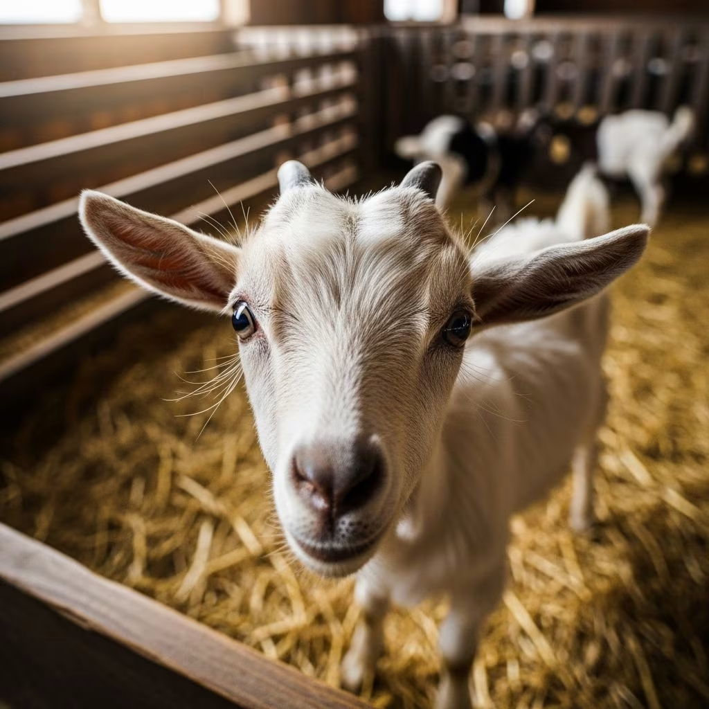
Coccidiosis is a prevalent intestinal disease in goats caused by protozoan parasites of the genus Eimeria. It primarily impacts young kids, leading to significant health challenges and economic losses in goat farming operations. This guide delves into the biology of the parasite, clinical manifestations, diagnostic approaches, management strategies, and therapeutic interventions to help goat owners safeguard their herds.
Understanding the Parasite Behind the Disease
The causative agents of coccidiosis in goats are various species of Eimeria, with Eimeria ninakohlyakimovae and Eimeria arloingi being among the most pathogenic. These single-celled parasites complete their life cycle within the intestinal cells of the host. Infection begins when goats ingest sporulated oocysts from contaminated environments, such as feces-laden pastures or bedding. Once inside the gut, oocysts release sporozoites that invade epithelial cells, multiply through schizogony and gametogony, and eventually produce new oocysts shed in feces.
Goat kids are particularly susceptible because their immune systems are immature. Adults typically develop immunity after early exposure, making clinical disease rare in mature animals. However, heavy environmental contamination can overwhelm even immune goats, especially under stress.
Risk Factors Fueling Outbreaks
Several farm management practices exacerbate coccidiosis risks. Overcrowding in pens increases oocyst transmission via direct fecal-oral contact. Wet, poorly drained areas promote oocyst sporulation, as these parasites require moisture and warmth (optimal at 25–30°C) to become infective within 1–3 days. Feeding on the ground or using low troughs allows fecal contamination of feed and water.
- High stocking densities in kidding areas
- Inadequate cleaning of bedding and facilities
- Stress from weaning, transport, or nutritional deficiencies
- Mixing age groups, exposing naive kids to oocysts from carriers
- Concurrent infections like worms or bacteria
Outbreaks peak in kids aged 5–8 weeks, coinciding with weaning when stress and dietary changes occur.
Recognizing Clinical Signs and Subclinical Effects
Symptoms vary from subclinical infections causing poor performance to acute, life-threatening disease. Subclinical cases manifest as reduced weight gain, unthrifty coats, and lower feed efficiency, often overlooked but contributing to long-term productivity losses.
Acute clinical coccidiosis presents dramatically:
- Watery or mucoid diarrhea, sometimes bloody or dark
- Straining (tenesmus) and rectal prolapse in severe cases
- Dehydration, sunken eyes, and weakness
- Anorexia, fever, and abdominal pain
- Rough hair coat, lethargy, and rapid emaciation
- Pasty feces progressing to severe dysentery
Without intervention, kids may become recumbent and die within days due to intestinal damage, blood loss, and secondary infections. Chronic cases lead to stunted growth, delayed puberty, and nutrient malabsorption from scarred intestines.
Diagnostic Methods for Accurate Confirmation
Suspected cases require fecal flotation to detect and quantify oocysts. A count exceeding 5,000–10,000 oocysts per gram indicates heavy infection warranting treatment, though clinical correlation is key as low counts can still cause disease in young kids.
| Method | Description | Advantages |
|---|---|---|
| Fecal Flotation | Centrifugal or sugar/salt solutions to concentrate oocysts | Simple, cost-effective; species identification possible |
| McMaster Technique | Quantifies oocysts per gram | Allows monitoring treatment efficacy |
| Post-Mortem Exam | Gut lesions: thickened intestines, hemorrhages | Confirms pathogenesis in fatalities |
| PCR Testing | Molecular detection of Eimeria DNA | High sensitivity for subclinical cases |
Differentiate from other diarrheas like enterotoxemia or bacterial infections via history and necropsy.
Prevention: Building a Resilient Herd Environment
Prevention hinges on breaking the life cycle through hygiene and strategic interventions. Maintain dry, clean pens with frequent bedding changes. Elevate feed and water to 2–3 feet off ground. Rotate pastures, allowing 3–6 months rest to reduce oocyst viability in cool/dry conditions.
Coccidiostats in feed prevent outbreaks:
- Decoquinate (Deccox): 0.5 mg/kg body weight; safe for all ages
- Lasalocid (Bovatec) or Monensin (Rumensin): 13–20 g/ton feed; toxic to horses/equines
Introduce gradually post-weaning. Immunity develops naturally; avoid over-sanitizing to allow controlled exposure. Vaccinate indirectly via managed challenge.
Treatment Protocols for Effective Recovery
Act swiftly upon symptoms: isolate cases, provide electrolytes for hydration, and administer supportive care like warmth and easily digestible feed. Veterinary-prescribed anticoccidials target parasite stages.
| Drug | Dosage | Duration | Notes |
|---|---|---|---|
| Amprolium (Corid) | 50 mg/kg/day PO or water | 5–7 days | Supplement thiamine (B1); inhibits rumen function |
| Sulfadimethoxine (Albon) | 55 mg/kg day 1, then 27.5 mg/kg | 4 days | Extra-label in goats; early use best |
| Sulfamethazine | 100 mg/kg PO q24h | 7 days | Effective in water; vet consult required |
| Toltrazuril/Ponazuril | Vet-determined | 1–2 doses | Targets multiple stages; emerging option |
Treat entire group if endemic. Monitor for resistance; rotate drugs. Severe cases may need fluids IV and pain relief.
Long-Term Management and Monitoring
Post-treatment, fecal oocyst counts should drop within 1–2 weeks. Implement biosecurity: quarantine new arrivals, avoid mixing ages. Nutritional support with probiotics aids gut recovery. Track growth rates and body condition scores quarterly.
For dairy goats, watch for hepatobiliary forms causing liver failure in adults.
FAQs on Coccidiosis in Goats
Q: Can adult goats get coccidiosis?
A: Rarely clinically, due to immunity, but they shed oocysts as carriers.
Q: Is Corid safe for goats?
A: Yes, with B1 supplementation; follow vet dosing.
Q: How to prevent in organic farms?
A: Hygiene, rotation, herbal supports like garlic; coccidiostats if allowed.
Q: What’s the prognosis with treatment?
A: Excellent if early; permanent damage possible if delayed.
Q: Do dewormers treat coccidiosis?
A: No; they target worms, not protozoa.
References
- Preventing and Treating Coccidiosis in Goats — Goat Journal. 2023. https://goatjournal.iamcountryside.com/health/coccidiosis-in-goats/
- Coccidiosis in Sheep and Goats — Kentucky Sheep and Goat. 2024. https://www.kysheepandgoat.org/post/coccidiosis-in-sheep-and-goats
- Coccidiosis in Goats and Sheep: Treatment and Prevention — Alabama Cooperative Extension System (ACES). 2023. https://www.aces.edu/blog/topics/animals-urban/coccidiosis-in-goats-and-sheep-treatment-and-prevention/
- Coccidiosis in a Goat Herd — Government of Manitoba (.gov). 2022. https://www.gov.mb.ca/agriculture/livestock/goat/coccidiosis-in-goats.html
- Management and treatment of coccidiosis in sheep and goats — Veterinary Times. 2024. https://www.vettimes.com/news/vets/livestock/management-and-treatment-of-coccidiosis-in-sheep-and-goats
- Observe young goats for possible coccidiosis symptoms — Michigan State University Extension (MSU). 2023. https://www.canr.msu.edu/news/observe_young_goats_for_possible_coccidiosis_symptoms
- Coccidiosis of Goats — Merck Veterinary Manual (Merck & Co.). 2025. https://www.merckvetmanual.com/digestive-system/coccidiosis/coccidiosis-of-goats



