

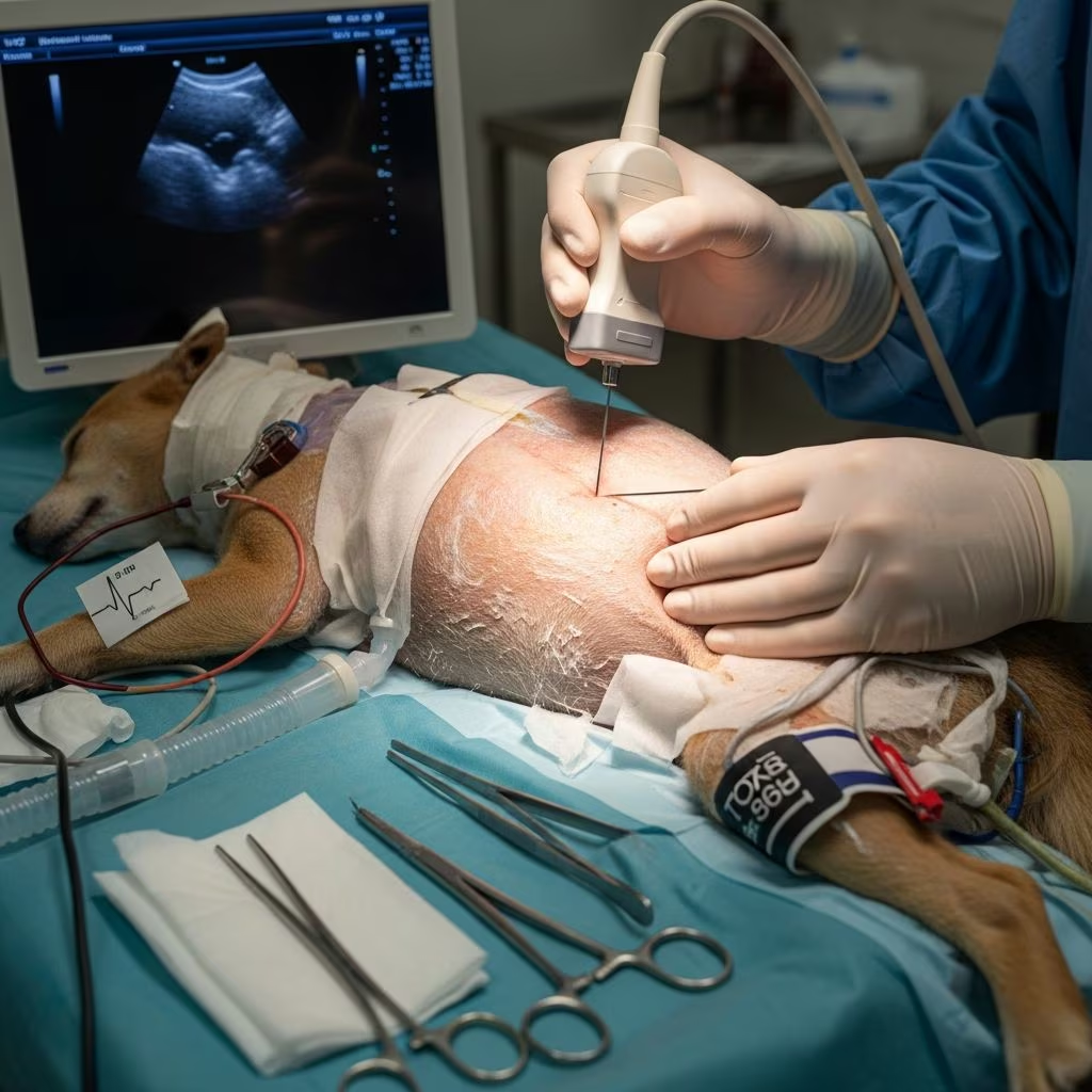
Gallbladder aspiration, known as cholecystocentesis, serves as a vital diagnostic tool in veterinary practice for small animals experiencing liver and biliary issues. This minimally invasive method allows veterinarians to collect bile samples directly from the gallbladder, enabling detailed analysis for infections, parasites, and other abnormalities. Primarily performed under ultrasound guidance, it offers a safer alternative to more invasive surgical approaches, particularly in dogs and cats showing signs of hepatobiliary disease.
Understanding the Role in Veterinary Diagnostics
In small animal medicine, liver disorders manifest through symptoms like jaundice, appetite loss, and elevated liver enzymes. Traditional diagnostics such as blood tests and imaging provide clues, but bile analysis via cholecystocentesis delivers definitive insights. This procedure is especially valuable for identifying bacterial cholangitis, parasitic infestations like Platynosomum in cats, and assessing gallbladder wall integrity.
The technique’s rise stems from its balance of efficacy and low risk. Studies confirm its utility in both healthy and diseased animals, with bile cultures and cytology revealing pathogens missed by fecal exams. For instance, in feline cases, bile egg counts surpass fecal detection sensitivity for platynosomiasis, making it indispensable for chronic cases.
Step-by-Step Procedural Guide
Performing cholecystocentesis requires precision, typically under sedation or general anesthesia to ensure patient comfort and stillness. Here’s a breakdown of the standard protocol:
- Pre-procedure Preparation: Withhold food for 12 hours while allowing water access. Conduct a full ultrasound to visualize the gallbladder, noting its position, size, and any wall thickening or sludge.
- Anesthesia and Positioning: Use propofol induction with isoflurane maintenance or heavy sedation. Position the animal in dorsal, left lateral, or ventral recumbency based on gallbladder accessibility, often through the ventral or ventrolateral liver parenchyma.
- Ultrasound Guidance: Employ a high-frequency linear transducer. Use color Doppler to avoid vessels. Clip and prepare the ventral abdomen aseptically.
- Needle Insertion: Advance a 22-gauge needle attached to an extension set, three-way stopcock, and 10 mL syringe through at least 2 cm of liver tissue into the gallbladder. Aspirate as much bile as possible to reduce leakage risk.
- Post-procedure Check: Re-scan for peritoneal fluid, hemorrhage, or gallbladder changes. Monitor packed cell volume (PCV) pre- and post-procedure.
This transhepatic approach minimizes bile peritonitis by sealing the puncture site naturally. Procedures often coincide with liver biopsies or other interventions, enhancing diagnostic yield without added sessions.
Safety Profile and Complication Rates
Safety data from multiple studies affirm cholecystocentesis as low-risk. In one cohort of 83 cats, 47% underwent general anesthesia, with minimal PCV changes (median -4%) despite concurrent fluids and procedures. Only one of five transfusions linked directly to hemorrhage.
Across dogs and cats, minor complications occur in 2.8-3.4% of cases, including transient gallbladder edema or mild peritoneal hemorrhage. No major bile leaks or fatalities reported in controlled settings. Post-procedure ultrasound confirms absence of free fluid in most instances.
| Species | Minor Complications (%) | Major Complications (%) | PCV Change (Median) |
|---|---|---|---|
| Cats | 2.8 | 0 | -4% |
| Dogs | 3.4 | <1 | Stable |
Factors like operator experience (board-certified radiologists) and complete gallbladder emptying mitigate risks. Even in inflamed gallbladders from cholangitis, safety holds, countering earlier concerns.
Diagnostic Applications and Bile Analysis
Bile samples yield cytology, cultures, and parasitology results. Cytological evaluation detects bacteria, inflammatory cells, and neoplasia. Aerobic/anaerobic cultures guide antibiotic therapy, crucial for ascending infections in cholangiohepatitis.
In cats with suspected Platynosomum, bile egg counts via stereomicroscopy offer higher sensitivity than fecal flotation. Drops as small as 10-20 μL enable rapid in-house screening. Abnormal ultrasound findings like wall thickening correlate loosely with infection; cats show 96% negative predictive value for normal scans.
- Bacterial Detection: Sensitivity 81% in dogs, higher NPV in cats.
- Parasitic Diagnosis: Superior to fecal tests for chronic platynosomiasis.
- Cytology Insights: Reveals sludge, crystals, or malignant cells from 140+ samples in combined dog/cat studies.
Dogs require culture regardless of ultrasound, given lower specificity. This complements liver biopsies for comprehensive hepatobiliary assessment.
Indications: When to Perform Cholecystocentesis
Indications include unexplained hyperbilirubinemia, persistent enzyme elevations, or ultrasound anomalies like distension or mucocele precursors. It’s ideal for:
- Chronic cholangitis/cholecystitis unresponsive to empiric therapy.
- Negative fecal parasitology with compatible signs.
- Pre-surgical planning for biliary diversion.
- Therapeutic decompression in select obstructive cases.
Avoid in coagulopathic patients without correction or inaccessible gallbladders. High NPV in feline gallbladder ultrasound supports selective use.
Comparing Techniques: PUC vs. Surgical Options
| Aspect | Percutaneous Ultrasound-Guided (PUC) | Laparoscopic/Surgical |
|---|---|---|
| Invasiveness | Minimally invasive | Moderately to highly invasive |
| Anesthesia | Sedation or light GA | Full GA |
| Complication Risk | Low (2-3%) | Higher (bile leak, hemorrhage) |
| Diagnostic Yield | High for culture/cytology | High + biopsy access |
| Recovery Time | Hours | Days |
PUC excels for outpatient diagnostics; surgery suits when biopsy or drainage needed.
Post-Procedure Care and Monitoring
Monitor for lethargy, vomiting, or abdominal pain 24-48 hours post-procedure. IV fluids mitigate hemodilution effects on PCV. Repeat bloodwork assesses anemia or ongoing inflammation. Most resume eating same day.
Advances and Future Directions
Fluoroscopic guidance emerges for drain placement in obstructions, expanding therapeutic roles. Ongoing cytology studies refine interpretation norms. Integration with advanced imaging promises refined patient selection.
Frequently Asked Questions (FAQs)
Is cholecystocentesis painful for my pet?
Under sedation/anesthesia, pets feel no pain. Mild discomfort may follow, managed with analgesics.
How accurate is bile culture for infections?
Highly accurate, especially cats (96% NPV); dogs need regardless of imaging.
Can it diagnose cancer?
Cytology suggests neoplasia; confirm with biopsy.
What’s the cost range?
Varies; ultrasound/sedation ~$500-1500 USD.
Is it safe for puppies/kittens?
Limited data; use cautiously, prefer experienced operators.
References
- Percutaneous cholecystocentesis in cats with suspected hepatobiliary disease — Vergauwen et al. 2024-05-15. https://pmc.ncbi.nlm.nih.gov/articles/PMC11104171/
- Percutaneous Ultrasound‐guided Cholecystocentesis and Bile Egg Counts in Cats with Platynosomum fastosum — Rosado et al. 2016-06-01. https://pmc.ncbi.nlm.nih.gov/articles/PMC4913578/
- Liver Biopsy and Cholecystocentesis — Veterian Key. 2016-01-01. https://veteriankey.com/liver-biopsy-and-cholecystocentesis/
- When should you do cholecystocentesis? — Veterinary View Box. 2024-01-01. https://veterinary-view-box.beehiiv.com/p/when-should-you-do-cholecystocentesis
- Cytological Findings of 140 Bile Samples from Dogs and Cats — RVC Repository. 2023-01-01. https://rvc-repository.worktribe.com/OutputFile/1398267



