

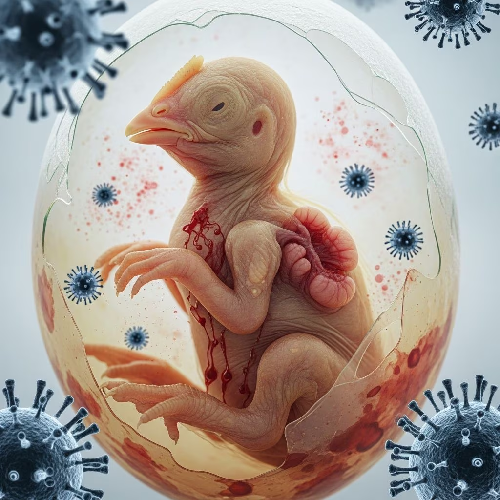
Chicken anemia virus (CAV), a highly contagious pathogen in the Anelloviridae family, poses significant challenges to global poultry production. This single-stranded DNA virus primarily targets young chicks, leading to severe anemia, stunted growth, and weakened immunity, while causing subtler but economically damaging effects in older birds. With its environmental resilience and dual vertical-horizontal transmission, CAV remains ubiquitous despite vaccination efforts.
The Nature and Biology of CAV
CAV belongs to the genus Gyrovirus and features a non-enveloped icosahedral structure, making it durable in litter, feces, and feathers. It replicates in hemocytoblasts of the bone marrow and precursor T cells in the thymus cortex, triggering apoptosis that depletes red blood cells, thrombocytes, and granulocytes. This dual assault results in anemia, hemorrhage risks, and broad immunosuppression, exacerbating secondary bacterial, viral, or fungal infections.
In molecular terms, CAV encodes three key proteins: VP1 (scaffolding and capsid), VP2 (cell binding), and VP3 (apoptosis inducer). These enable efficient infection of dividing cells, with peak viremia occurring 3-4 days post-infection in 1-day-old chicks. Recent genomic studies reveal strain diversity, influencing virulence; for instance, certain isolates correlate with outbreaks in layer hens.
Global Spread and Transmission Dynamics
CAV infects poultry flocks worldwide, thriving in intensive operations due to its stability outside hosts. Horizontal spread occurs via fecal-oral contact, contaminated litter, feather dander, and possibly aerosols. Vertical transmission happens when unexposed hens lay viremic eggs or roosters shed virus in semen, infecting progeny before maternal antibodies develop.
- Fecal-oral route: Primary in broilers, amplified by crowded housing.
- Feather transmission: Virus persists in follicle epithelium, aiding flock-wide dissemination.
- Vertical pathway: Critical in breeder flocks lacking prior immunity, leading to 100% infection rates in naive hatchmates.
Subclinical infections in breeders perpetuate cycles, with waning maternal antibodies leaving 2-4 week-old chicks vulnerable. Co-infections with pathogens like infectious bursal disease virus heighten pathogenicity and transmissibility.
Recognizing Clinical Manifestations in Young Chicks
Outbreaks strike chicks aged 7-14 days lacking maternal protection, peaking at 2-3 weeks. Classic signs include lethargy, pallor, ruffled feathers, and hemorrhages in skin, muscles, and viscera. Hematocrit plummets to 6-27%, with mortality reaching 10-50% during acute phases 5-6 days post-onset.
| Age Group | Key Symptoms | Mortality Risk |
|---|---|---|
| 1-2 weeks | Pale comb, weakness, subcutaneous bleeding | High (up to 55%) |
| 2-4 weeks | Anemia, poor weight gain, thymic atrophy | Moderate (5-10%) |
| >4 weeks | Subclinical; secondary infections | Low |
Mortality declines after 10-12 days as survivors recover, but survivors exhibit lasting growth deficits and vaccine response failures.
Subclinical Impacts on Adult Flocks and Economics
In birds over 4 weeks, CAV rarely causes overt disease but induces subclinical immunosuppression, reducing feed efficiency, body weight, and slaughter yields. This manifests as higher condemnation rates and vulnerability to gangrenous dermatitis, inclusion body hepatitis, or hydropericardium syndromes when co-occurring with other agents.
Economic tolls include diminished broiler performance and layer productivity losses, amplified in unvaccinated operations. Studies link seropositive flocks to 5-10% revenue drops from suboptimal gains and increased secondary disease burdens.
Pathological Changes and Lesion Identification
Necropsy reveals pale bone marrow (yellowish), thymic cortex depletion of lymphoblasts, splenic hypocellularity, and occasional myocardial pallor. Bone marrow aplasia confirms erythroid/myeloid progenitor loss, while thymic atrophy underscores T-cell destruction. These lesions, absent in healthy chicks, guide presumptive diagnosis amid clinical signs.
Advanced Diagnostic Approaches
Live-bird confirmation relies on hematocrit <27% paired with PCR detection of CAV DNA in blood lymphocytes. Postmortem, immunohistochemistry identifies antigens in thymus/bone marrow, complemented by quantitative PCR for viremia levels. Serology via ELISA detects antibodies but cannot distinguish active from past infections.
- PCR: Gold standard for sensitivity/specificity; detects as low as 10 viral copies.
- Viral isolation: Rare due to fastidious growth in cell culture (MDCC-MSB1 line).
- Serology: Monitors flock immunity post-vaccination.
Differentiate from mareks, IBD, or reticuloendotheliosis via multiplex PCR panels.
Management Strategies Without Specific Therapeutics
No antiviral treatments exist; supportive care focuses on warmth, electrolytes, and antibiotics for secondary bacteria. Control hinges on breeder immunity: vaccinate hens pre-lay (live attenuated strains at 14-18 weeks) to confer maternal antibodies protecting chicks 2-3 weeks.
Breeder exposure pre-production builds lifelong immunity, halting vertical spread. Biosecurity minimizes horizontal risks: all-in-all-out systems, litter management, and disinfection (though CAV resists many agents). Monitor via sentinel serology.
Vaccination Protocols and Efficacy
Live CAV vaccines (e.g., Cux-1 strain) induce sterilizing immunity in breeders without egg transmission risks post-seroconversion. Administer subcutaneously or intraocularly; efficacy exceeds 95% in preventing clinical chick disease. In ovo vaccination trials show promise for broilers.
| Vaccine Type | Target Flock | Dose/Route | Protection Duration |
|---|---|---|---|
| Live attenuated | Breeders (14-18 wk) | SC/IO | Lifelong; maternal to chicks |
| In ovo | Broilers (18 days incub.) | Injection | 3-4 weeks |
Emerging Challenges and Research Directions
Strain variability challenges universal vaccines; genomic surveillance tracks virulent clades linked to outbreaks in Asia/Europe. Human gyrovirus homologs raise zoonotic queries, though avian strains remain chicken-specific. Future foci: recombinant subunit vaccines and antivirals targeting VP3 apoptosis.
Frequently Asked Questions (FAQs)
What is the most effective way to prevent CAV in poultry?
Vaccinating breeders before egg production ensures maternal antibody transfer to chicks, blocking vertical transmission.
Can CAV infect other species besides chickens?
Primarily chickens; experimental infections occur in turkeys, but natural hosts are avian poultry.
How long does CAV survive in the farm environment?
Highly stable; persists months in litter/feces, resisting standard disinfectants.
Is CAV infection curable once symptoms appear?
No specific cure; survivors recover but face growth penalties. Prevention is key.
What secondary diseases does CAV predispose flocks to?
Gangrenous dermatitis, IBH, E. coli infections due to neutropenia and lymphopenia.
References
- Chicken Anemia Virus Infection — Merck Veterinary Manual. 2023. https://www.merckvetmanual.com/poultry/chicken-anemia-virus-infection/chicken-anemia-virus-infection
- Chicken Infectious Anemia Virus: Immunosuppressive Diseases — Mansoura Veterinary Medical Journal. 2023-10-01. https://mvmj.researchcommons.org/cgi/viewcontent.cgi?article=1250&context=home
- Chicken anemia virus — Wikipedia (primary sources referenced). 2023. https://en.wikipedia.org/wiki/Chicken_anemia_virus
- Isolation, Identification, and Whole Genome Analysis of Chicken Anemia Virus — PMC/NCBI. 2023-07-20. https://pmc.ncbi.nlm.nih.gov/articles/PMC10386238/
- Genetic diversity, distribution, and evolution of chicken anemia virus — Frontiers in Microbiology. 2023-10-10. https://www.frontiersin.org/journals/microbiology/articles/10.3389/fmicb.2023.1145225/full



