

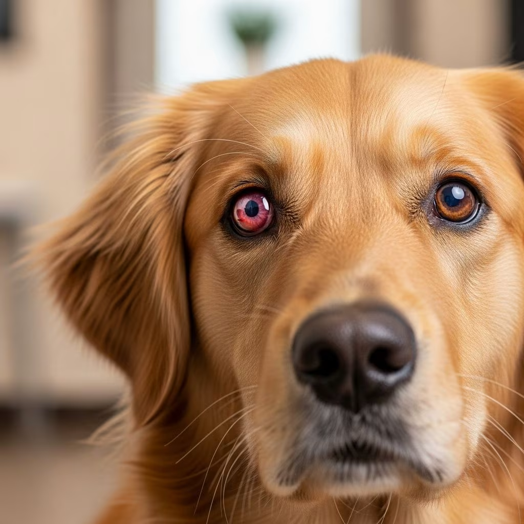
Cherry eye in dogs is a noticeable condition where the tear-producing gland from the dog’s third eyelid prolapses, appearing as a red, fleshy mass in the corner of the eye. While not immediately painful or vision-threatening, it requires prompt veterinary attention to prevent complications like dry eye syndrome, which can lead to chronic discomfort and potential vision loss.
What is Cherry Eye in Dogs?
Dogs possess a third eyelid, also known as the nictitating membrane, located in the inner corner of the eye. This structure includes a tear gland responsible for producing up to 30-50% of the eye’s tear film, essential for lubrication and eye health. Cherry eye occurs when the fibrous attachment holding this gland in place weakens, causing it to prolapse or ‘pop out’. The exposed gland becomes swollen, inflamed, and bright red—resembling a cherry, hence the name.
This condition is not cancerous or infectious but a mechanical issue stemming from anatomical weakness. Unlike in humans, a dog’s third eyelid serves critical functions, including tear production and protection during activities like hunting or swimming. When prolapsed, the gland cannot function properly, leading to inadequate lubrication and increased risk of secondary issues.
Symptoms of Cherry Eye in Dogs
Recognizing cherry eye early is key to effective management. The primary symptom is a **red, pink, or fleshy mass** protruding from the medial (inner) corner of the eye, often smooth and triangular-shaped. This mass may intermittently retract but typically persists or worsens over time.
- Pawing or rubbing at the eye: Dogs often scratch or rub the affected eye due to discomfort, potentially worsening irritation.
- Squinting or frequent blinking: Indicates pain or foreign body sensation from the prolapse.
- Redness and inflammation: The sclera (white of the eye) may appear bloodshot; the mass itself swells and reddens.
- Mucus discharge or dryness: Reduced tear production leads to thick discharge, crusting, or dry eye signs like dull cornea.
- Swelling around the eye: Mild eyelid swelling may accompany the prolapse.
These symptoms can mimic allergies, infections, or foreign bodies, so professional diagnosis is essential. If untreated, the gland may fibrose (harden), impairing function permanently.
Causes of Cherry Eye in Dogs
The root cause is a **weak or absent fibrous attachment** securing the third eyelid gland to the eye’s lower rim. This allows prolapse during excitement, straining, or normal movement.
Key contributing factors include:
- Genetics: Hereditary weakness in connective tissue is primary. A 2022 study of over 900,000 dogs confirmed higher incidence in purebreds, especially young ones under 1 year.
- Breed predisposition: Brachycephalic (flat-faced) and certain spaniel breeds are most affected due to anatomical traits.
- Age: Typically seen in puppies and young dogs under 2 years, during rapid growth.
- Trauma or inflammation: Eye injuries, allergies, or irritants can weaken tissues further.
- Cartilage abnormalities: In large breeds like Great Danes, scrolled third eyelid cartilage contributes.
Environmental factors like allergies exacerbate swelling, preventing natural repositioning.
Breeds Prone to Cherry Eye
Certain breeds have a significantly higher risk due to genetic and conformational factors. Brachycephalic breeds suffer from crowded facial structures weakening gland support.
| Breed Group | Common Breeds | Risk Factors |
|---|---|---|
| Brachycephalic | English Bulldog, French Bulldog, Pug, Boston Terrier, Shih Tzu | Flat face compresses eye structures |
| Spaniels & Hounds | Cocker Spaniel, Beagle, Bloodhound, Lhasa Apso | Loose connective tissue |
| Large Breeds | Great Dane, Mastiff, Saint Bernard | Cartilage scrolling |
| Other | Neapolitan Mastiff, Newfoundland | Genetic predisposition |
Mixed breeds have lower odds, per large-scale studies. Owners of at-risk breeds should monitor young pups closely.
Diagnosis of Cherry Eye
Veterinarians diagnose cherry eye via physical exam, noting the characteristic prolapse. Fluorescein staining rules out ulcers, while tear production tests (Schirmer tear test) assess gland function—critical as the gland produces 30-50% of tears.
Advanced imaging or referral to a veterinary ophthalmologist may evaluate cartilage or chronic damage. Differential diagnoses include tumors, allergies, or prolapsed conjunctiva.
Treatment Options for Cherry Eye in Dogs
Early treatment preserves gland function and prevents dry eye (keratoconjunctivitis sicca, KCS). Surgery is the gold standard, with over 90% success rates.
- Manual repositioning: Temporary for mild, recent cases; gland pushed back, often with meds.
- Surgical pocket technique (imbrication): Most common—creates a pocket to secure gland with sutures, preserving function.
- Anti-inflammatories: Steroids or NSAIDs reduce swelling pre/post-surgery.
- Artificial tears/lubricants: Supportive for moisture during recovery.
- Gland removal (muclectomy): Last resort for non-viable glands; risks lifelong KCS meds.
Surgery is outpatient, with 10-14 day recovery: e-collar, drops, restricted activity. Recurrence is low with proper technique.
Recovery and Aftercare
Post-op, monitor for infection, suture reaction, or recurrence. Administer meds as prescribed, prevent rubbing. Full recovery takes 2-4 weeks; regular tear tests ensure no KCS.
Long-term: At-risk breeds may need bilateral monitoring, as the other eye often follows.
Complications if Untreated
Prolonged prolapse damages the gland, causing dry eye—corneal ulcers, pigmentation, vision loss. Secondary infections or chronic pain ensue.
Prevention Tips
- Early socialization and allergy management in pups.
- Regular vet eye exams for predisposed breeds.
- Avoid trauma; supervise play.
Breeder screening for hereditary weakness reduces incidence.
Frequently Asked Questions (FAQs)
What is cherry eye in dogs?
A prolapse of the third eyelid’s tear gland, appearing as a red mass in the eye corner.
Is cherry eye painful for dogs?
Not acutely, but causes irritation leading to rubbing and discomfort.
Will cherry eye resolve without surgery?
Rarely; it typically persists or recurs, risking complications.
How much does cherry eye surgery cost?
Varies by location/clinic; consult your vet for estimates (not specified in sources).
Can cherry eye affect both eyes?
Yes, often sequentially in predisposed dogs.
What breeds get cherry eye most?
Bulldogs, Cocker Spaniels, Shih Tzus, Pugs.
References
- What is Cherry Eye in Dogs? | AZ Vet Direct — AZ Vet Direct. 2023. https://azvet.direct/low-cost-pet-surgery-in-mesa-and-the-entire-southeast-valley-of-phoenix/what-is-cherry-eye-in-dogs-6-symptoms-you-should-watch-out-for/
- Cherry Eye in Dogs — VCA Animal Hospitals. 2024. https://vcahospitals.com/know-your-pet/cherry-eye-in-dogs
- Understanding Cherry Eye In Pets: Causes, Symptoms, And Treatment — Village Oaks Veterinary Center. 2023. https://vovcbham.com/blog/understanding-cherry-eye-in-pets-causes-symptoms-and-treatment/
- Cherry Eye in Dogs: What Does It Look Like, and Why Does It Happen? — PetMD. 2024. https://www.petmd.com/dog/conditions/eyes/cherry-eye-in-dogs
- Cherry Eye in Dogs — Blue Cross. 2023. https://www.bluecross.org.uk/advice/dog/health-and-injuries/cherry-eye-in-dogs
- Cherry Eye in Dogs: Causes, Symptoms & Treatment — Pawlicy Advisor. 2024. https://www.pawlicy.com/blog/cherry-eye-in-dogs/
- Cherry Eye in Dogs: Signs, Symptoms, Treatments — American Kennel Club. 2024. https://www.akc.org/expert-advice/health/cherry-eye-in-dogs/



