

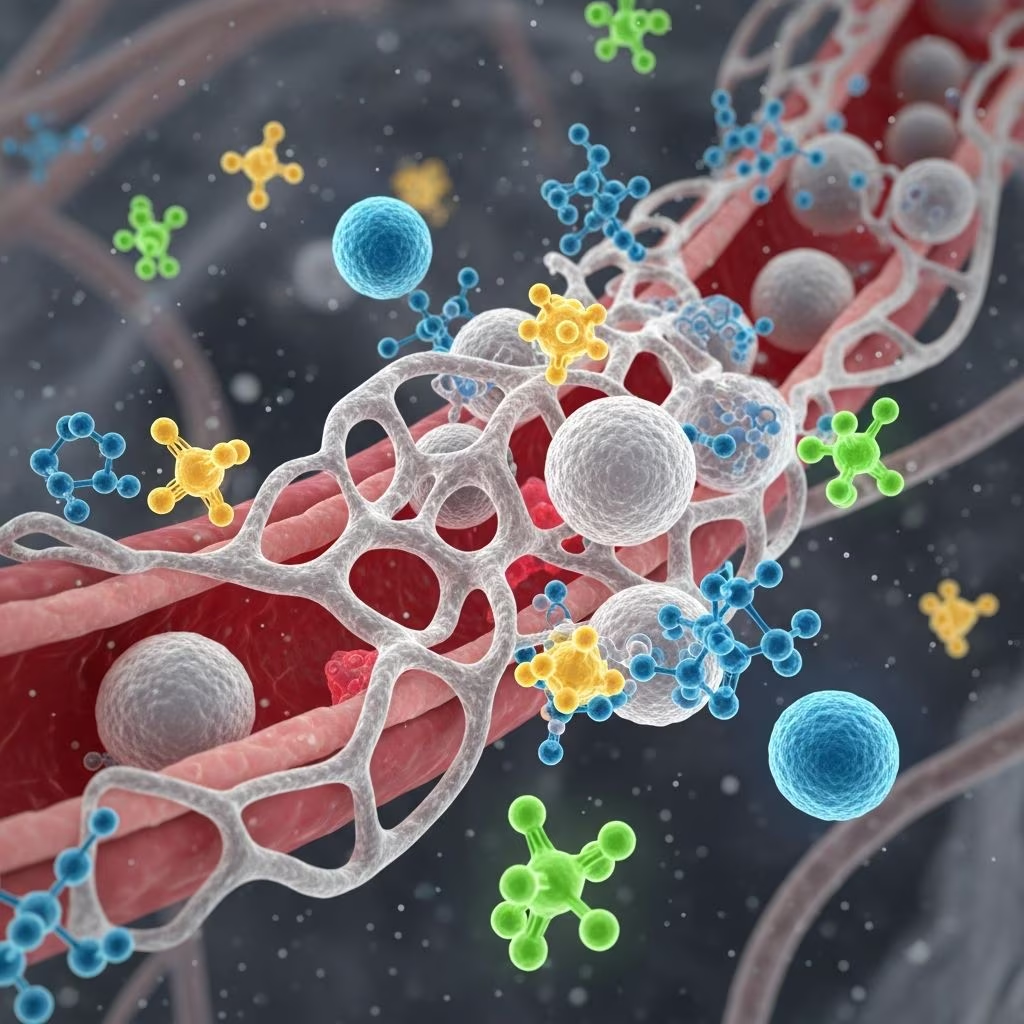
Inflammation serves as a fundamental defense mechanism in animals, triggered by injury, infection, or irritants. Central to this process are chemical mediators—molecules released from cells, plasma, and damaged tissues that coordinate vascular alterations, immune cell recruitment, and tissue repair. These agents amplify the response to eliminate threats while promoting recovery, though dysregulation can lead to chronic conditions.
Foundational Role of Inflammatory Mediators
The inflammatory cascade begins with tissue damage prompting the liberation of mediators from resident cells like mast cells and infiltrating leukocytes. These substances induce vasodilation, increase vascular permeability, and attract white blood cells, forming the hallmarks of acute inflammation: redness, heat, swelling, and pain. Vasoactive amines initiate rapid changes, while slower-acting lipids and proteins sustain the response.
Understanding these mediators is crucial in veterinary medicine, as they underpin treatments like NSAIDs targeting eicosanoids or antihistamines blocking amines. Historical experiments, such as those demonstrating histamine’s role in skin flare reactions, laid the groundwork for modern pharmacology.
Primary Vasoactive Amines and Their Rapid Effects
**Histamine and serotonin** stand out as the quickest-acting mediators, stored pre-formed in mast cells and platelets. Histamine binds H1 receptors on endothelial cells, causing immediate arteriolar dilation and gap formation between vessels, allowing plasma leakage. This exudate dilutes toxins and delivers antibodies and complement proteins to the site.
Serotonin, prominent in rodents and some ruminants, complements histamine by further contracting venular sphincters, enhancing leakage. Both amines provoke pain by stimulating nerve endings and contract smooth muscle, contributing to symptoms like bronchospasm in allergic reactions.
- Histamine sources: Mast cells, basophils, enterochromaffin cells.
- Triggers: Physical injury, C3a/C5a anaphylatoxins, IgE cross-linking.
- Duration: Effects peak within minutes and fade as amines are metabolized by histaminase or diluted.
Peptide Mediators: Kinins and Complement Fragments
Peptides like **bradykinin**, generated via the kinin system from plasma precursors, powerfully dilate vessels and boost permeability while directly activating pain fibers. Unlike amines, kinins arise from enzymatic cleavage during clotting and complement activation, linking coagulation to inflammation.
Complement fragments **C3a and C5a** (anaphylatoxins) degranulate mast cells, release histamine, and act as potent chemoattractants for neutrophils. C5a further promotes lysosomal enzyme release from leukocytes, amplifying tissue degradation to access pathogens. These peptides bridge innate immunity and inflammation.
| Mediator | Main Action | Cell Targets |
|---|---|---|
| Bradykinin | Vasodilation, pain, permeability | Endothelium, nerves |
| C5a | Chemotaxis, degranulation, enzyme release | Neutrophils, mast cells |
| C3a | Mast cell activation, smooth muscle contraction | Mast cells, vessels |
Lipid-Derived Eicosanoids: Prostaglandins, Leukotrienes, and More
Arachidonic acid metabolites, or eicosanoids, derive from membrane phospholipids via cyclooxygenase (COX) and lipoxygenase pathways, offering sustained inflammatory modulation. **Prostaglandins (PGs)** like PGE2 and PGI2, produced by COX-1 and COX-2, sensitize pain receptors and promote fever via hypothalamic effects while vasodilating.
Genetic studies in mice reveal COX-1’s underappreciated role in early inflammation; COX-1 knockouts show reduced paw swelling in models, challenging the COX-2-only paradigm. **Leukotrienes (LTs)**, especially LTB4, excel in chemotaxis, drawing neutrophils potently, while LTC4/D4 cause prolonged constriction in airways and vessels. Thromboxane A2 (TXA2) from platelets aggregates cells and constricts vessels, counterbalancing prostacyclin’s dilation.
Platelet-activating factor (PAF), another lipid, mirrors LTs in chemotaxis and activates endothelium for leukocyte adhesion.
Cytokines: Orchestrators of Immune Amplification
**Cytokines** such as TNF-α, IL-1β, IL-6, and IL-8 are polypeptides from macrophages and lymphocytes that systemically and locally regulate inflammation. TNF-α and IL-1β induce endothelial adhesion molecules (e.g., E-selectin), enabling leukocyte rolling and extravasation. They also trigger acute-phase protein synthesis in the liver.
In endotoxemia models, LPS from Gram-negative bacteria spikes TNF-α, IL-6, and IL-1β, causing toxicity, fever, and shock in species like cattle, pigs, and mice. IL-8 specifically chemoattracts neutrophils, while IL-10 provides counter-regulation to prevent excess damage.
- Pro-inflammatory: TNF-α (endothelial activation, cachexia), IL-1β (fever, ACTH release), IL-6 (acute-phase induction).
- Chemotactic: IL-8, MIP-2.
- Anti-inflammatory: IL-10, TGF-β.
Acute-Phase Proteins: Systemic Response to Inflammation
The acute-phase response, driven by IL-6 and glucocorticoids, elevates liver-produced proteins by 1.5-5 fold. **C-reactive protein (CRP)** opsonizes bacteria for phagocytosis. **Serum amyloid A (SAA)** modulates chemokines and HDL function. **Haptoglobin** binds free hemoglobin, sequestering iron from bacteria, while **fibrinogen** aids clotting and leukocyte margination.
In challenge studies, LPS elicits the highest rises in haptoglobin and SAA compared to whole bacteria or outer membrane proteins, linking to severe tissue injury and neuroinflammation. These proteins mark inflammation severity and predict outcomes in infections.
| Protein | Function | Inflammatory Stimulus Example |
|---|---|---|
| Haptoglobin | Hemoglobin binding, bacteriostasis | LPS inoculation |
| SAA | Chemoattractant, lipid transport | Gram-negative sepsis |
| Fibrinogen | Clotting, cell adhesion | Tissue trauma |
Leukocyte Contributions and Phagocytosis Signaling
Neutrophils and monocytes release mediators post-extravasation. Activated monocytes produce TNF-α, IL-1β, IL-6, NO, and chemokines like MCP-1 during phagocytosis. Phagocytosis of opsonized particles involves Rho GTPases, PI3K, and phospholipase C, fusing lysosomes with phagosomes.
These cells amplify inflammation via reactive oxygen/nitrogen species and enzymes, but also resolve it through apoptosis and efferocytosis.
Therapeutic Targeting of Mediators in Veterinary Practice
Glucocorticoids suppress phospholipase A2, blocking eicosanoid synthesis. NSAIDs inhibit COX, reducing PGs. Mast cell stabilizers prevent amine release. Cytokine blockers like anti-TNF antibodies treat autoimmune diseases in animals. Balancing inhibition avoids immunosuppression.
Common Questions on Inflammatory Mediators
What triggers the release of chemical mediators in animals?
Tissue injury, pathogens (e.g., LPS), or immune complexes activate resident cells and plasma systems.
How do eicosanoids differ from vasoactive amines?
Amines act pre-formed and rapidly; eicosanoids are synthesized on-demand, sustaining effects.
Why are cytokines critical in systemic inflammation?
They coordinate leukocyte trafficking, fever, and acute-phase responses across organs.
Can acute-phase proteins diagnose inflammation?
Yes, elevations in haptoglobin or SAA indicate ongoing inflammation, especially post-infection.
What role does COX-1 play versus COX-2?
Both contribute; COX-1 initiates acute responses, per knockout studies.
References
- The crucial roles of inflammatory mediators in inflammation: A review — T. M. T. Nguyen, A. A. A. Saleh, S. A. H. Al-Suhaibani. 2018-05-25. https://pmc.ncbi.nlm.nih.gov/articles/PMC5993766/
- The crucial roles of inflammatory mediators in inflammation: A review [PDF] — T. M. T. Nguyen et al. 2018. https://pdfs.semanticscholar.org/c4c0/85b91129edc49f9335054e0ebec664e887a7.pdf
- Inflammation and Healing — Veterian Key. Accessed 2026. https://veteriankey.com/inflammation-and-healing/
- The crucial roles of inflammatory mediators in inflammation: A review — Veterinary World. 2018-05-30. https://www.veterinaryworld.org/Vol.11/May-2018/9.html
- Actions of Inflammatory Mediators Table — Merck Veterinary Manual. Accessed 2026. https://www.merckvetmanual.com/multimedia/table/actions-of-inflammatory-mediators
- Pathophysiology of Inflammation in Animals — MSD Veterinary Manual. Accessed 2026. https://www.msdvetmanual.com/pharmacology/inflammation/pathophysiology-of-inflammation-in-animals



