

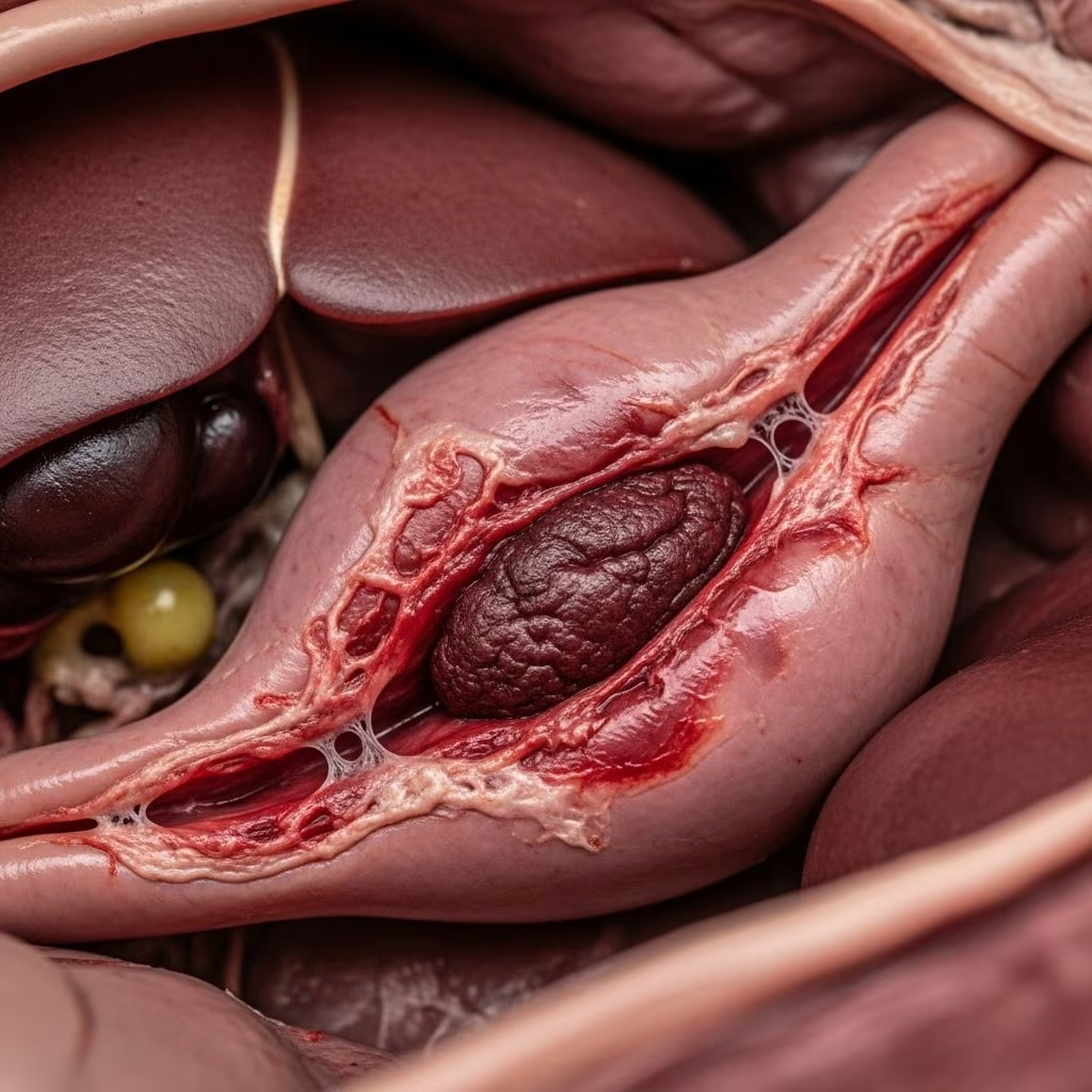
Caudal vena cava thrombosis represents a severe and often lethal vascular disorder in cattle, primarily affecting animals in intensive production systems like feedlots and high-yield dairy operations. This condition arises when septic thrombi form in the posterior vena cava, typically due to liver abscesses eroding into the vessel, leading to embolic spread and destructive pneumonia throughout the lungs. Despite advances in veterinary care, the prognosis remains dismal, with mortality approaching 100%, underscoring the need for proactive prevention through optimized feeding practices.
The Pathophysiological Cascade: From Feed to Fatal Clots
The journey to caudal vena cava thrombosis often begins in the rumen. High-grain diets, common in finishing cattle, disrupt rumen pH balance, causing subacute ruminal acidosis. This acidity damages the rumen wall, fostering bacterial invasion and rumenitis. Pathogens migrate to the liver via the portal vein, forming hepatic abscesses. When these abscesses penetrate the adjacent caudal vena cava, bacteria-laden thrombi develop. Fragments break off, traveling to the pulmonary arteries, where they lodge and spawn multifocal abscesses, aneurysms, and hemorrhages.
This hematogenous dissemination distinguishes the disease from primary respiratory infections. Unlike bacterial pneumonias confined to lung lobes, lesions here are diffuse, reflecting embolic origins. Supporting evidence from necropsy studies consistently reveals thrombi occluding the vena cava, with hepatic lesions in nearly all cases, confirming the liver’s central role.
- Ruminal trigger: Acidosis from rapid grain adaptation erodes rumen mucosa.
- Portal spread: Bacteria reach liver, forming abscesses in 10-30% of feedlot cattle on high-concentrate feeds.
- Vascular invasion: Abscesses erode vena cava wall, seeding septic clots.
- Embolic phase: Thrombi fragment, embolizing to lungs via right heart.
Recognizing the Signs: Acute and Chronic Presentations
Clinical manifestations vary from sudden collapse to insidious decline, complicating early detection. Acute cases strike rapidly, with cattle exhibiting severe respiratory distress, open-mouth breathing, and frothy hemoptysis—bright red blood coughed from ruptured pulmonary aneurysms. Tachypnea exceeds 60 breaths per minute, accompanied by tachycardia over 100 bpm, pale mucosae, and harsh lung sounds.
Chronic forms mimic wasting diseases, featuring progressive emaciation, intermittent fever, chronic cough, and melena (digested blood in feces). Affected animals weaken over weeks to months, showing exercise intolerance, ventral edema, and bottle-jaw in advanced stages. Heart murmurs from anemia or right-sided strain are common. In dairy cows, milk drop signals systemic involvement.
| Phase | Key Symptoms | Duration |
|---|---|---|
| Acute | Hemoptysis, dyspnea, collapse | Days to 1 week |
| Chronic | Weight loss, cough, fever | Weeks to months |
| Terminal | Sternal recumbency, shock | Hours |
Differential diagnoses include traumatic reticuloperitonitis, pulmonary thromboembolism from other sources, or hardware disease, but hemoptysis and multifocal crackles point uniquely to embolic pneumonia.
Diagnostic Approaches: Confirming the Unseen Clot
Ante-mortem diagnosis relies on history and exam, bolstered by imaging. Ultrasonography reveals vena caval thrombi as hypoechoic masses with acoustic shadowing, often with hepatic abscesses visible cranially. Thoracic ultrasound shows pleural effusion and pulmonary consolidations. Radiography, though less sensitive in large animals, may depict aneurysms or abscess shadows.
Bloodwork discloses anemia (PCV <20%), leukocytosis, and hyperfibrinogenemia, reflecting chronic inflammation. Fecal occult blood tests positive in melena cases. Definitive confirmation comes postmortem: vena cava contains firm, septic thrombus; lungs harbor myriad abscesses (0.5-3 cm), aneurysms, and clots. Hepatic abscesses, often Trueperella pyogenes-positive, adjoin the vessel.
Treatment Realities: Limited Options and Ethical Choices
Therapeutic success is rare due to entrenched infection and vascular occlusion. Antimicrobials like penicillin or cephalosporins target Fusobacterium and Arcanobacterium but fail to resolve established thrombi. Supportive measures—flunixin meglumine for pain, fluids for dehydration, NSAIDs for inflammation—offer palliation but not cure. Thrombolytics or surgery, viable in small animals, are impractical and risky in cattle.
Given the guarded-to-grave prognosis, euthanasia or salvage slaughter (if regulations permit and welfare allows) is recommended upon strong suspicion. Economic losses from death or cull compound the toll.
Prevention Strategies: Breaking the Acidosis-Liver-Clot Chain
Control hinges on averting ruminal acidosis, the root cause. Gradual diet transitions over 3-4 weeks when stepping up grains minimize pH drops. Ionophores like monensin buffer rumen acidity, slashing abscess incidence by 40-50%. Balanced rations with 10-20% roughage sustain fiber mat integrity.
- Feeding protocols: Increase concentrates by <10% weekly; provide ad lib hay.
- Additives: Monensin (200-300 mg/head/day) reduces acidosis risk.
- Monitoring: Track manure consistency; soft pellets signal acidosis.
- Herd health: Cull chronic offenders; vaccinate against rumen pathogens if available.
In outbreak herds, screen via liver condemnations at slaughter; adjust protocols accordingly. These measures can drop thrombosis rates below 1%.
Economic and Welfare Impacts on Producers
Beyond individual losses, caudal vena cava thrombosis erodes profitability. Feedlot mortality hits 2-5% in unmanaged groups, with carcass condemnations for abscesses costing $50-100/head. Dairy impacts include lost lactation (up to 20% yield drop) and culling primes prematurely.
Welfare concerns amplify: protracted suffering from dyspnea and pain demands swift intervention. Producers must weigh humane endpoints against salvage value, guided by AVMA euthanasia guidelines.
Research Frontiers and Future Directions
Ongoing studies probe genomics for acidosis susceptibility and novel antimicrobials for abscess penetration. Biomarkers like serum haptoglobin may enable early screening. Precision feeding via rumen sensors promises real-time pH monitoring, potentially revolutionizing prevention.
Frequently Asked Questions (FAQs)
What causes blood clots in cattle vena cava?
Primarily liver abscesses from ruminal acidosis erode the vessel, forming septic thrombi.
Is there a cure for vena cava thrombosis in cows?
No effective cure exists; supportive care rarely succeeds, and euthanasia is often necessary.
How do you prevent this in feedlots?
Gradual grain adaptation, ionophores, and roughage inclusion are key.
What does hemoptysis look like in affected cattle?
Frothy, bright red blood from mouth/nose, signaling ruptured lung aneurysms.
Can ultrasound diagnose this condition?
Yes, it visualizes vena caval thrombi and liver abscesses reliably.
References
- Vena Caval Thrombosis and Metastatic Pneumonia in Cattle — Merck Veterinary Manual. 2023. https://www.merckvetmanual.com/respiratory-system/non-infectious-respiratory-system-diseases-in-cattle/vena-caval-thrombosis-and-metastatic-pneumonia-in-cattle
- Thrombosis of Vena Cava — NADIS. 2022. https://www.nadis.org.uk/disease-a-z/cattle/thrombosis-of-vena-cava/
- Thrombosis, Embolism, Aneurysm, and Dissection in Animals — MSD Veterinary Manual. 2024. https://www.msdvetmanual.com/circulatory-system/thrombosis-embolism-aneurysm-and-dissection/thrombosis-embolism-aneurysm-and-dissection-in-animals
- Caudal vena cava thrombosis in Cows (Bovis) — Vetlexicon. 2023. https://www.vetlexicon.com/bovis/alimentary/articles/caudal-vena-cava-thrombosis/
- EP 76: Vena Caval Thrombosis in Cattle — Pet Care Partner (YouTube). 2023-10-15. https://www.youtube.com/watch?v=ZhVG-hs0C1M
- Caudal vena caval thrombosis following treatment of deep digital septicitis in a cow — Canadian Veterinary Journal (PMC). 2011-12-01. https://pmc.ncbi.nlm.nih.gov/articles/PMC3258833/



