

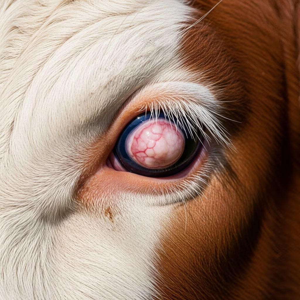
Eye tumors represent a significant health concern in cattle operations worldwide, affecting productivity and animal welfare. The most prevalent eye neoplasms affecting cattle include squamous cell carcinoma and lymphosarcoma with orbital involvement. Understanding these conditions, their development patterns, and management strategies is essential for veterinarians and livestock producers committed to maintaining herd health and identifying affected animals early.
Understanding the Most Common Ocular Neoplasms in Cattle
The primary tumor types affecting cattle eyes are squamous cell carcinoma (SCC) and lymphosarcoma. Squamous cell carcinoma represents the most frequently encountered ocular neoplasm in cattle, typically developing in the outer layer of cells covering the eye and surrounding structures. Lymphosarcoma, when affecting the orbital region, causes progressive bilateral eye protrusion, reduced eye movement capacity, exposure-related corneal inflammation, and ulcerations that may progress to corneal rupture if left untreated.
The distinction between these tumor types is clinically important because they present differently and require different management approaches. Squamous cell carcinoma typically progresses slowly through identifiable stages, while lymphosarcoma develops more rapidly and affects the structures surrounding the eye rather than the visible surface.
Environmental and Genetic Risk Factors Contributing to Tumor Development
Tumor development in cattle eyes is multifactorial, with ultraviolet radiation serving as one of the most significant environmental contributors. Cattle exposed to high solar radiation in areas with extended daylight hours or at high altitudes experience increased risk. The cumulative nature of UV exposure means that older animals generally have higher incidence rates than younger stock.
Pigmentation status emerges as the most critical genetic factor influencing tumor development. Cattle lacking pigmentation around the eyelids and on the conjunctiva face substantially elevated risk compared to pigmented individuals. This explains why breeds such as Herefords with white facial markings and light-colored hair around the eyes show higher prevalence rates. Unpigmented tissue lacks protective melanin that shields cells from UV-induced DNA damage.
Nutritional plane also contributes to tumor development risk. High-quality nutrition, while beneficial for overall cattle productivity, paradoxically increases tumor occurrence, suggesting that metabolic factors associated with enhanced cell division may facilitate neoplastic transformation.
Anatomical Locations and Frequency of Tumor Presentation
Ocular tumors in cattle demonstrate preferences for specific anatomical sites. The limbal region, representing the junction between the cornea and sclera, represents the most frequently affected location. The third eyelid, upper eyelid margin, and lower eyelid margin also commonly develop tumors. Bilateral involvement, where tumors develop in both eyes, occurs in approximately 35 percent of cases, necessitating comprehensive examination of both eyes during diagnostic assessment.
The distribution pattern reflects differential exposure to solar radiation and variations in protective mechanisms across different ocular structures. The limbal region, being fully exposed and less protected by eyelid closure mechanisms, receives maximal radiation exposure.
Stages of Squamous Cell Carcinoma Progression
Squamous cell carcinoma progresses through four distinct developmental stages, each presenting different clinical and pathologic characteristics:
- Stage One—Plaques: Initially, small circular white elevations appear on the conjunctival surface. These lesions appear benign and smooth. Approximately 30 percent of plaques may spontaneously regress without intervention, representing the only stage with potential for natural resolution.
- Stage Two—Keratomas: Hard, raised growths develop on eyelid surfaces, typically coated with ocular secretions and cellular debris. These lesions represent progressive transformation but remain technically benign.
- Stage Three—Papillomas: Wart-like growths emerge, presenting as irregular raised tissue. These lesions still lack malignant potential but indicate advancing disease progression.
- Stage Four—Carcinomas: Malignant transformation produces nodular, cauliflower-like masses. These lesions are friable, often bloody, ulcerated, and characteristically foul-smelling due to necrosis and secondary bacterial infection. Malignant carcinomas possess the ability to invade adjacent and underlying tissues, including the eyeball and orbital structures.
Not all carcinomas develop through these sequential stages. Some may bypass benign precursor lesions and appear directly as malignant masses, highlighting the variable biologic behavior of these neoplasms.
Clinical Recognition and Diagnostic Features
Early-stage eye tumors may prove difficult to identify, particularly when limited to the third eyelid where they appear as small, knobby growths barely visible on casual observation. Initial signs include mild eye discharge resulting from tissue irritation and slight discomfort.
As tumors enlarge, clinical signs become more apparent:
- Progressive elevation and irregularity of lesion margins
- Transition from white or grey coloration to pink or red appearance
- Increased ocular discharge, often serosanguineous in nature
- Hemorrhage from the tumor surface
- Necrosis manifesting as blackened, sloughing tissue
- Secondary fly attraction and pestering behavior
- Foul odor from advanced necrotic lesions
Advanced tumors with significant size may impair vision or cause mechanical irritation, affecting grazing behavior and overall performance. Invasion into deeper orbital tissues results in exophthalmos (eye protrusion), reduced ocular mobility, and exposure-related corneal complications.
Lymph node involvement becomes apparent when parotid and submandibular nodes around the head and neck enlarge, indicating metastatic disease progression.
Lesion Characteristics by Anatomical Location
Tumors originating on corneal and conjunctival surfaces typically begin at the limbal sclera and gradually extend across the cornea. Lesions in this location often progress from benign smooth white plaques through intermediate stages to squamous cell carcinoma, though direct malignant transformation without obvious precursor lesions may occur.
Eyelid-based lesions present differently, typically beginning as either ulcerative lesions or hyperkeratotic growths resembling cutaneous horns. These lesions may develop less predictably through the staged progression seen with corneoscleral tumors.
Tumors may enlarge substantially without invading the globe itself; however, in advanced disease, invasion into the eye and orbit becomes inevitable, with spread to regional lymph nodes representing late-stage metastatic disease.
Diagnostic Confirmation and Differential Considerations
Clinical diagnosis typically relies on characteristic appearance of lesions during ophthalmologic examination. However, definitive diagnosis requires biopsy and histologic examination to confirm neoplastic changes and distinguish neoplasia from other conditions.
Important differential diagnoses to exclude include severely traumatized eyes with disorganized tissue architecture and advanced infectious keratoconjunctivitis with extensive corneal involvement. These conditions may superficially resemble advanced ocular malignancy but carry different prognostic implications and treatment requirements.
Impact on Cattle Productivity and Welfare
Ocular neoplasia significantly affects affected animals’ quality of life and economic productivity. Tumors causing vision impairment alter grazing behavior, reduce feed intake, and compromise performance metrics. Animals with bilateral eye involvement may experience temporary or permanent blindness, creating welfare concerns and safety hazards within the herd environment.
The slow development of many tumors means affected cattle may remain productive during early stages, but as disease progresses, declining performance becomes inevitable. The expense of managing affected animals must be weighed against remaining productive life expectancy and overall herd economics.
Management and Treatment Approaches
Management strategies vary depending on disease stage, tumor location, and individual animal circumstances. Early benign lesions may be monitored for spontaneous regression, particularly plaques that demonstrate potential for natural resolution.
Surgical intervention offers the most definitive treatment approach for tumors limited to specific locations. Lesions confined to the third eyelid may be surgically removed under local anesthesia. Cryosurgery represents an alternative technique using controlled freezing to destroy tumor tissue.
Advanced cases involving the eyeball itself may require enucleation (eye removal) under standing sedation with local anesthetic blocks. However, this approach may not be economically justifiable for commercial cattle destined for processing, particularly when welfare considerations necessitate culling rather than attempted preservation.
The presence of orbital invasion, regional lymph node involvement, or bilateral disease substantially complicates treatment feasibility and reduces likelihood of successful outcomes, often necessitating humane culling decisions based on animal welfare considerations.
Prevention Strategies and Risk Reduction
Effective prevention focuses on selecting genetically resistant animals and managing environmental exposure. Breeding cattle with pigmented skin surrounding the eyelids represents the most practical long-term prevention strategy. Genetic selection for increased periocular pigmentation reduces individual susceptibility regardless of management practices.
Environmental management strategies include providing shade structures during periods of intense solar radiation and managing grazing rotations to limit UV exposure during peak radiation hours. These measures prove particularly valuable in high-altitude regions or areas with extensive daylight periods.
Regular visual inspection of animals, particularly during routine handling and management activities, enables early identification of developing lesions when intervention options remain most favorable.
Breed and Individual Susceptibility Variations
Certain breeds demonstrate higher disease prevalence due to genetic predisposition toward reduced periocular pigmentation. Hereford cattle with characteristic white facial blazes and light coloration around the eyes exemplify this vulnerability. Conversely, breeds maintaining darker pigmentation patterns show substantially lower disease incidence.
Individual animals within susceptible breeds vary in disease development, suggesting additional genetic factors beyond simple pigmentation status influence tumor susceptibility. Age represents another critical factor, with older animals showing higher incidence reflecting cumulative UV exposure over extended lifespans.
Frequently Asked Questions About Cattle Eye Tumors
How quickly do eye tumors progress in cattle?
Progression speed varies substantially among individual animals and tumor types. Early benign lesions may remain static for extended periods, while malignant carcinomas can progress from small lesions to large masses over months. Some plaques spontaneously regress, while others progress directly to carcinoma without obvious intermediate stages.
Can eye tumors in cattle be prevented entirely?
Complete prevention is not possible, but risk reduction is achievable through selective breeding for increased periocular pigmentation and environmental management minimizing UV exposure. Genetic selection represents the most effective long-term strategy.
What is the prognosis for cattle with diagnosed eye tumors?
Prognosis depends on tumor stage, location, and extent of tissue involvement. Early benign lesions carry favorable prognosis for management, while advanced malignant lesions with orbital invasion carry poor prognosis, typically resulting in culling decisions based on welfare and economic considerations.
Are there medical treatments for cattle eye tumors?
No systemic medical treatments exist for ocular neoplasia in cattle. Management relies on surgical intervention for accessible lesions or culling for advanced disease. Early detection enables more treatment options before extensive tissue involvement develops.
References
- Eye Conditions in Cattle — NADIS (National Animal Disease Information System). 2024. https://www.nadis.org.uk/disease-a-z/cattle/eye-conditions-in-cattle/
- Ocular Neoplasia in Cattle — Merck Veterinary Manual. 2024. https://www.merckvetmanual.com/eye-diseases-and-disorders/neoplasia-of-the-eye-and-associated-structures/ocular-neoplasia-in-cattle
- Squamous Cell Carcinoma of the Bovine Eye — Kansas State University College of Veterinary Medicine. 2024. https://www.vet.k-state.edu/docs/vhc/farm/ag-practice-updates/Bovine_Cancer_Eye.pdf
- Eye Cancer in Cattle — Agriculture Victoria Livestock Health and Welfare. 2024. https://agriculture.vic.gov.au/livestock-and-animals/livestock-health-and-welfare/eye-cancer-in-cattle
- Catching Cancer Eye Early — VetSouth. 2024. https://www.vetsouth.co.nz/blog/post/126141/catching-cancer-eye-early/
- Investigation of DNA Damage and Apoptosis in Bovine Ocular Squamous Cell Carcinoma — PubMed Central. 2024. https://pmc.ncbi.nlm.nih.gov/articles/PMC12199710/
- Keep an Eye on the Eyes: Veterinary Viewpoints — Oklahoma State University College of Veterinary Medicine. 2024. https://news.okstate.edu/articles/veterinary-medicine/2024/keep-an-eye-on-the-eyes.html



