

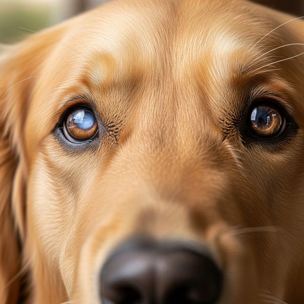
Understanding Cataracts in Dogs
Cataracts are a common eye condition that affects dogs of all ages, breeds, and sexes. When a dog develops a cataract, the normally clear lens becomes cloudy or opaque, interfering with light reaching the retina. This cloudiness can significantly impact your dog’s vision and quality of life. While cataracts may start small and barely noticeable, they often grow progressively, potentially leading to severe vision impairment or blindness if left untreated. Understanding this condition is essential for dog owners, as early detection and intervention can help preserve your pet’s eyesight and overall well-being.
What Causes Cataracts in Dogs?
Cataracts develop in dogs for various reasons, and understanding the underlying causes can help you better protect your pet’s eye health.
Primary Causes of Cataracts
Hereditary and Genetic Factors: The most common cause of cataracts in dogs is hereditary or genetic disease. Certain dog breeds have a genetic predisposition to develop cataracts, including American Cocker Spaniels, Labrador Retrievers, French Poodles, Boston Terriers, Bichon Frises, and Welsh Springer Spaniels. If you own one of these breeds, genetic testing may be available to identify carriers of genes linked to hereditary cataract formation.
Age-Related Cataracts: As dogs age, they may lose the ability to restore old or damaged lens protein fibers as quickly as they did in their youth. This natural aging process can lead to cataract development in senior dogs.
Diabetes: Cataracts occur commonly as a complication of diabetes in dogs. High blood sugar levels can cause changes in the lens that lead to cataract formation.
Other Contributing Factors: Trauma to the eye, inflammation, nutritional deficiencies, and certain medications can also contribute to cataract development in some dogs.
Recognizing the Signs of Cataracts in Dogs
The most obvious sign of cataracts in dogs is a change in the appearance of the eye. Owners often notice a cloudy or opaque appearance to the cornea, which may look milky-white or grayish. At certain angles or lighting conditions, the affected lens may sparkle or display a crystalline effect. However, physical appearance is just one indicator of this condition.
Behavioral Signs to Watch For
Beyond the visible cloudiness, your dog may exhibit behavioral changes that suggest vision loss:
– Increased clumsiness or bumping into objects, especially in low-light conditions- Reluctance to jump, climb stairs, or navigate unfamiliar spaces- Changes in activity level or reluctance to go outside- Excessive blinking or eye discomfort- Squinting or keeping eyes partially closed- Anxiety or hesitation when moving around
Dogs with cataracts experience vision similar to looking through frosted glass, which can be disorienting and frustrating for your pet. Even though cataracts may initially be small and not interfere with vision, they often grow and can drastically impair eyesight over time.
Stages of Cataracts in Dogs
Cataracts develop progressively through distinct stages, and understanding which stage your dog’s cataracts have reached is crucial for determining the appropriate treatment approach.
The Four Stages of Canine Cataracts
| Stage | Lens Coverage | Symptoms | Vision Impact |
|---|---|---|---|
| Incipient | Less than 15% | Pinpoint cloudiness, often unnoticeable | No significant vision loss |
| Immature | 15-99% | Increasing cloudiness and opacity | Variable vision impairment |
| Mature | 100% | Complete lens opacity | Near-blindness or total blindness |
| Hypermature | 100% (advanced) | Lens material breakdown, potential complications | Total blindness with increased risk of secondary issues |
It’s important to note that cataracts don’t always progress through all four stages. Some dogs’ cataracts may stabilize at a particular stage and progress no further. Additionally, your dog may have different stages of cataracts in each eye or even a cataract in only one eye.
Diagnosing Cataracts in Dogs
Proper diagnosis is essential for determining the best course of treatment for your dog’s cataracts. A comprehensive diagnostic approach helps your veterinarian assess the severity, identify underlying causes, and determine whether your dog is a candidate for surgical intervention.
Initial Examination
When you bring your dog to the veterinarian, they will begin by taking a detailed medical history and asking about when you first noticed the symptoms. Your vet will perform a complete physical examination, paying special attention to your dog’s eyes and the structures surrounding them. Initial diagnostic testing, such as blood work and urinalysis, usually doesn’t show abnormalities unless there is co-existing disease, such as diabetes or hypocalcemia.
Specialized Diagnostic Tests
Slit-Lamp Biomicroscopy: This specialized examination uses a focused beam of light to allow direct visualization and assessment of the lens. The slit lamp provides magnification and illumination, enabling your veterinarian to evaluate the cloudiness and condition of the lens in detail.
Schirmer Tear Test: A small filter paper is placed inside the dog’s lower eyelid to measure tear production. This test helps rule out dry eye as a contributing factor to eye problems.
Ocular Ultrasound: This imaging technique is particularly helpful when advanced cataracts make visualization of the retina difficult through the cloudy lens. The ultrasound allows your veterinarian to assess the back of the eye and check for conditions such as retinal detachment or lens rupture.
Electroretinography (ERG): This test measures the electrical activity of the retina in response to light stimulation. An ERG is crucial for confirming that your dog’s retina is functioning properly before cataract surgery is considered. This test helps ensure that vision restoration is possible after surgical intervention.
Treatment Options for Cataracts in Dogs
Treatment approaches for canine cataracts depend on the stage of the cataract, your dog’s overall health, and whether your dog is a suitable candidate for surgery.
Non-Surgical Management
For early-stage or incipient cataracts that don’t significantly affect vision, your veterinarian may recommend monitoring the condition rather than pursuing immediate treatment. Regular eye examinations and careful observation of your dog’s vision and behavior are essential during this phase. While non-surgical treatments such as eye drops exist, they typically cannot reverse cataract formation or stop progression entirely.
Surgical Treatment
Surgery is the most effective and long-lasting solution for cataracts in dogs, particularly when performed before the cataract reaches full maturity and before secondary inflammation develops. Approximately 95% of dogs successfully regain vision following cataract surgery when performed by an experienced veterinary ophthalmologist.
Understanding Cataract Surgery in Dogs
Pre-Surgery Evaluation
Before proceeding with cataract surgery, your dog must undergo comprehensive pre-operative testing to ensure they are a suitable candidate and can safely undergo general anesthesia.
Blood Work: Screening blood work is performed to ensure your dog is healthy enough to undergo general anesthesia safely.
Pre-Surgical Ultrasound: An ocular ultrasound is conducted to assess the back of your pet’s eye, since advanced cataracts make direct visualization difficult. This imaging helps detect issues such as retinal detachment or lens rupture.
Electroretinogram (ERG): As mentioned earlier, an ERG measures the retina’s electrical activity in response to light stimulus, confirming your dog’s retina is healthy and can process light after surgery. If these pre-surgical tests reveal unexpected issues such as retinal problems, your dog may unfortunately not be suitable for cataract surgery.
The Surgical Procedure
Cataract surgery in dogs is performed under general anesthesia, with a muscle relaxant administered to help the eye sit in the correct position for the operation. Once your dog is properly anesthetized, the veterinary ophthalmologist makes a small incision into the eye.
The surgeon then uses a special instrument that employs ultrasonic waves to break the diseased lens into small fragments through a process called phacoemulsification. This same technique is used in human cataract surgery. The veterinary ophthalmologist continues this process until all lens fragments have been surgically removed.
Following lens removal, an artificial lens implant, known as an intraocular lens (IOL), is implanted into the eye. This step is critically important because without a lens to focus light onto the retina, your dog’s vision would remain very blurry. The artificial lens allows images to be focused clearly onto the retina, restoring functional vision.
Recovery and Aftercare Following Cataract Surgery
Successful recovery from cataract surgery requires careful attention to your veterinarian’s post-operative instructions. The initial healing period following cataract surgery typically lasts approximately two weeks, during which your dog requires significant care and monitoring.
Post-Surgery Care Requirements
During the recovery period, your dog must:
– Wear an Elizabethan collar (cone) at all times to prevent rubbing or scratching the eye- Have activity strictly restricted to leash walks only, with no running, jumping, or rough play- Receive multiple medications including prescription eye drops and oral medications as prescribed by your veterinarian- Attend all scheduled follow-up appointments for monitoring and assessment- Have their eye examined regularly to ensure proper healing
Carefully following your veterinarian’s instructions during this recovery period is absolutely essential for achieving a good outcome and preserving your dog’s restored vision. Any deviation from post-operative protocols could compromise the surgery’s success and your dog’s long-term vision.
Potential Complications and Long-Term Considerations
While cataract surgery is generally successful, it’s important to be aware of potential complications. Untreated cataracts can lead to secondary complications including uveitis (inflammation of the uveal tract), glaucoma (increased intraocular pressure), and lens luxation (dislocation of the lens), all of which are typically painful and can further compromise vision.
For dogs in which cataract surgery is not performed, continued monitoring remains very important. Regular veterinary check-ups help detect and manage any complications early. Your veterinarian will provide recommendations for ongoing treatment and monitoring to maintain your dog’s comfort and eye health.
Frequently Asked Questions About Dog Cataracts
Q: Can cataracts be prevented in dogs?
A: While hereditary cataracts cannot be prevented, maintaining good nutrition, managing underlying conditions like diabetes, and avoiding eye trauma can help reduce the risk in some cases. Regular veterinary eye exams are important for early detection.
Q: How quickly do cataracts progress in dogs?
A: Cataract progression varies significantly between individual dogs and causes. Some cataracts may remain stable for years, while others progress rapidly to blindness within months. Regular monitoring helps track progression in your specific dog.
Q: Is cataract surgery painful for dogs?
A: No, cataract surgery is performed under general anesthesia, so your dog will not experience pain during the procedure. Post-operative discomfort is typically minimal when proper pain management and aftercare are provided.
Q: Can cataracts develop in the other eye after surgery?
A: Yes, if cataracts are hereditary or age-related, they can develop in the other eye. Many dogs eventually require surgery on both eyes if they are suitable candidates.
Q: How long do artificial lens implants last in dogs?
A: Artificial lens implants are designed to be permanent. When properly placed and cared for, they should remain functional throughout your dog’s life, providing lasting vision restoration.
References
- Cataracts in Dogs: Signs, Causes, and Treatment — PetMD. 2024. https://www.petmd.com/dog/conditions/eye/c_dg_cataract
- Cataracts in Dogs — VCA Animal Hospitals. 2024. https://vcahospitals.com/know-your-pet/cataracts-in-dogs
- Disorders of the Lens in Dogs — Merck Veterinary Manual. 2024. https://www.merckvetmanual.com/dog-owners/eye-disorders-of-dogs/disorders-of-the-lens-in-dogs
- Comprehensive Guide to Cataract Surgery for Dogs — Pacific Santa Cruz Veterinary Hospital. 2023-10-15. https://www.pacificsantacruzvet.com/site/blog/2023/10/15/dog-cataract-surgery
- Cloudy Vision, Clear Solutions: A Pet Owner’s Guide to Cataracts — Veterinary Vision Center. 2024. https://veterinaryvisioncenter.com/cloudy-vision-clear-solutions-a-pet-owners-guide-to-cataracts/
- Cataracts in Dogs: A Comprehensive Guide — The Whole Pet Vet. 2024. https://toegrips.com/cataracts-in-dogs/
- Cataracts—A Guide for Pet Owners — VRC Malvern. 2024. https://www.vrcmalvern.com/cataracts-guide-for-pet-owners/



