

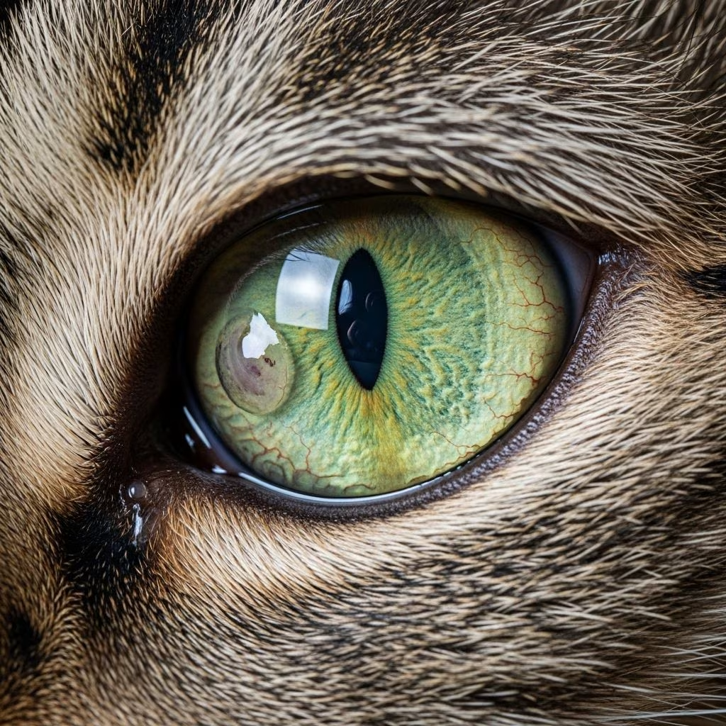
Corneal ulcers represent a serious threat to feline eye health, manifesting as erosions or defects in the cornea—the clear outer layer of the eye. These injuries can range from superficial scratches to deep perforations, potentially leading to vision loss if not addressed promptly. Cats frequently develop these ulcers due to viral infections, trauma, or anatomical issues, making early detection crucial for effective management.
Understanding the Cornea and Ulcer Formation
The cornea serves as the eye’s protective dome, maintaining transparency while shielding internal structures. In cats, its vulnerability stems from constant exposure and thin epithelial layers. Ulcers form when this epithelium erodes, exposing underlying stroma to irritants, bacteria, or viruses. Superficial ulcers affect only the surface, while deeper stromal ones penetrate further, increasing perforation risks.
Factors compromising corneal integrity include dry eye conditions, where reduced tear production fails to lubricate and protect. Eyelid abnormalities, such as entropion—where lids roll inward—can chronically abrade the surface. Foreign bodies like plant fibers or dust exacerbate damage, particularly in active outdoor cats.
Primary Culprits Behind Feline Corneal Ulcers
Feline herpesvirus type 1 (FHV-1) tops the list of causes, responsible for many chronic or recurrent cases. This virus triggers dendritic ulcers—branching patterns resembling tree limbs—distinguishing them from trauma-induced linear scratches. FHV-1 persists lifelong in carriers, reactivating under stress, illness, or immunosuppression.
Trauma accounts for acute incidents, often from clawing playmates, rubbing against rough surfaces, or chemical exposures. Bacterial superinfections complicate matters, delaying healing. Less common triggers involve fungal invasions in immunocompromised cats or breed-specific issues like brachycephalic faces in Persians, promoting exposure keratitis.
- Viral origins: Predominantly FHV-1, leading to ulcerative keratitis.
- Mechanical trauma: Scratches from fights or self-trauma.
- Anatomical defects: Eyelid malpositions or lagophthalmos.
- Environmental factors: Dryness, irritants, or foreign debris.
Spotting the Warning Signs Early
Cats mask discomfort adeptly, but corneal ulcers betray themselves through distinct behaviors. Excessive blinking or squinting signals irritation, as affected eyes become hypersensitive to light—photophobia. Profuse tearing or mucoid discharge accumulates, often staining fur beneath the eye. Cats paw at faces relentlessly, risking further injury.
Corneal cloudiness emerges as haze or opacity over the lesion, contrasting the normally crystal-clear surface. In severe cases, a black or pigmented spot called a corneal sequestrum appears, indicating necrosis. Vision impairment shows as bumping into objects or dilated pupils. Prompt veterinary consultation prevents progression to globe rupture.
| Symptom | Description | Severity Indicator |
|---|---|---|
| Squinting | Partial eye closure due to pain | Mild to severe |
| Discharge | Watery or pus-like from eye | Infection present |
| Cloudy cornea | Hazy or white appearance | Deep ulcer |
| Pawing/rubbing | Frequent face scratching | High discomfort |
| Sequestrum | Dark spot on cornea | Advanced necrosis |
Diagnostic Approaches for Accurate Assessment
Veterinarians employ fluorescein staining as the gold standard: a dye highlights defects under cobalt blue light, revealing ulcer depth and shape. Schirmer tear tests quantify lubrication deficits. Tonometry measures intraocular pressure, ruling out glaucoma mimics.
Cytology swipes identify bacterial or viral culprits, guiding antimicrobials. PCR tests confirm FHV-1 presence. Ultrasound probes deeper non-perforating ulcers when opacity obscures views. For sequestra, biopsy differentiates from neoplasia.
Medical Management Strategies
Treatment prioritizes pain relief, infection control, and epithelial regeneration. Broad-spectrum antibiotic drops or ointments—such as fluoroquinolones or triple antibiotic combos—combat bacteria every 4-6 hours initially. Antivirals like trifluridine or ganciclovir target FHV-1 topically; systemic famciclovir suits stressed or non-compliant cats.
Atropine or cycloplegics ease ciliary spasm and uveitis, though monitoring tears prevents iatrogenic dryness. Serum therapy, using cat’s own blood-derived drops, supplies growth factors for healing. Pain modulators like oral NSAIDs reduce inflammation without steroids, contraindicated in ulcers due to delayed repair.
- Antibiotics: Prevent secondary invasion.
- Antivirals: Inhibit FHV-1 replication.
- Cycloplegics: Relieve spasms.
- Regeneratives: Promote tissue regrowth.
Surgical Interventions for Refractory Cases
Non-healing ulcers exceeding 50% stromal depth demand surgery. Superficial debridement removes loose tissue, followed by third-eyelid flaps or nictitating membrane grafts for protection. Deep defects require corneal transplants or amniotic membrane overlays.
Sequestrectomy excises necrotic cores, often with beta-irradiation or grafts. Grid keratotomy—superficial scoring—stimulates healing in indolent ulcers. Referral to veterinary ophthalmologists ensures specialized tools like operating microscopes.
Home Care Essentials for Optimal Recovery
Elizabethan collars (e-cones) prevent self-trauma indispensably. Administer drops meticulously: one at a time, massaging lids gently. Clean discharges with saline-soaked pads, avoiding tap water. Indoor confinement minimizes re-injury risks.
Nutritional support via lysine supplements curbs FHV-1 in prone cats. Stress reduction through pheromones or quiet spaces aids immunity. Frequent vet rechecks—every 3-7 days—track fluorescein clearance.
- Secure cone collar continuously.
- Apply medications on schedule.
- Gently cleanse eye daily.
- Confine indoors fully.
- Monitor for worsening signs.
Preventing Recurrence in At-Risk Cats
Vaccination against FHV-1 forms the cornerstone, though not fully preventive in carriers. Routine lysine (250-500mg daily) inhibits viral shedding. Address dry eye proactively with cyclosporine ointments. Trim claws and supervise multi-cat interactions.
Breeds like Persians benefit from surgical eyelid corrections. Annual ophthalmic exams catch subclinical issues. Stress management—consistent routines, Feliway diffusers—keeps latent viruses dormant.
Prognosis and Long-Term Outlook
Superficial ulcers heal in 5-7 days with compliance, boasting 90% success. Deep or herpetic ones extend to weeks, with 70-80% vision preservation. Sequestra post-surgery recur less than 20%. Blindness risks rise with perforation or scarring.
Factors favoring recovery: early intervention, antiviral use, no comorbidities. Chronic FHV-1 cats thrive on maintenance therapy, maintaining quality vision.
Frequently Asked Questions
Can corneal ulcers heal without treatment?
Minor abrasions may self-resolve in days, but most feline cases, especially herpetic, worsen without intervention, risking perforation.
How often should I apply eye drops?
Typically every 4-6 hours, tapering as healing progresses; follow vet schedules precisely.
Is surgery always needed?
No, only for deep, non-healing, or sequestrum cases; medical therapy suffices for 80%.
Why does my cat keep getting ulcers?
Recurrent FHV-1 carriage, dry eye, or conformational issues; diagnostics pinpoint causes.
Can I use human eye drops?
Never; formulations differ, and some delay healing or cause toxicity.
Emerging Therapies on the Horizon
Regenerative drops harnessing stem cell factors accelerate repair. Corneal cross-linking fortifies collagen against melting. Gene therapies targeting FHV-1 latency promise breakthroughs, currently in trials.
References
- Treatment of Corneal Ulcers: What are the Medical Options? — PMC/NCBI. 2024. https://pmc.ncbi.nlm.nih.gov/articles/PMC11318758/
- Cat Corneal Ulcer: Causes, Treatments, and FAQs — PetMD. 2023-09-15. https://www.petmd.com/cat/conditions/eyes/c_ct_corneal_ulcers
- Corneal Ulcers — Cornell University College of Veterinary Medicine. 2022. https://www.vet.cornell.edu/departments-centers-and-institutes/cornell-feline-health-center/health-information/feline-health-topics/corneal-ulcers
- Corneal ulcers – Veterinary Teaching Hospital — Colorado State University. 2024. https://vetmedbiosci.colostate.edu/vth/services/ophthalmology/corneal-ulcers/
- Understanding and Managing Eye Ulcers in Cats — Horner Veterinary Specialists. 2023-11-20. https://hornervets.com/newsletter-blog/eye-ulcers-in-cats



