

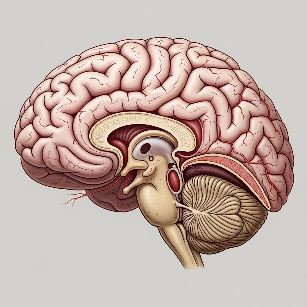
The brain of a domestic cat is a compact yet highly efficient organ, typically weighing around 25-30 grams in an average adult feline, with a cerebral cortex surface area of approximately 83 cm². This structure enables cats to exhibit extraordinary agility, sharp senses, and complex behaviors despite their small stature.
Physical Dimensions of the Feline Brain
A cat’s brain measures roughly 5-6 cm in length, 3-4 cm in width, and 2-3 cm in height, fitting snugly within the cranium. Unlike the smoother lissencephalic brains of smaller mammals, cat brains are gyrencephalic, featuring folded surfaces that increase surface area for enhanced processing power. High-resolution MRI studies at 3 Tesla reveal over 119 distinct structures, including fine details like cranial nerves and cerebellar nuclei previously hard to visualize.
Key measurements include:
- Cerebral cortex surface area: ~83 cm² (about 13 square inches)
- Cortical neurons: Approximately 763 million
- Visual cortex neuron density: 51,400 per mm³
- Cerebellum weight: 0.17% of total body weight
These specs highlight why cats excel in visual acuity and motor precision.
Core Divisions of the Cat Brain
The feline central nervous system centers on the brain, divided into three primary sections: the cerebrum for higher cognition, the cerebellum for coordination, and the brainstem for vital functions. This tripartite design mirrors human brain organization but is scaled down for a predator’s needs.
Cerebrum: Hub of Cognition and Senses
Dominating the brain’s front, the cerebrum handles decision-making, memory, and sensory integration. It comprises two hemispheres linked by the corpus callosum, facilitating inter-hemisphere communication and motor control. The caudate nucleus within supports learning, social bonding, and reward processing, explaining cats’ selective affections and trainability.
Cerebellum: Master of Balance and Movement
Positioned rearward, the cerebellum fine-tunes posture, gait, and reflexes. It enables the famous ‘righting reflex,’ allowing mid-air adjustments to land on feet. MRI imaging clearly delineates cerebellar folia, nuclei like dentate and fastigial, and pathways such as the spinocerebellar tract.
Brainstem: Regulator of Essentials
At the base, the brainstem connects to the spinal cord, originating 12 cranial nerve pairs for senses and facial movements. The medulla oblongata here modulates heart rate, respiration, and blood pressure, ensuring survival.
Detailed Structures Enhancing Feline Abilities
Beyond primaries, secondary components amplify cat prowess. Cranial nerves, especially the trigeminal, relay facial sensations. The olfactory bulb, prominent in cats, processes scents crucial for hunting and territory marking.
| Structure | Location | Primary Function |
|---|---|---|
| Cerebral Hemispheres | Forebrain | Information integration, voluntary movement |
| Corpus Callosum | Midline | Hemispheric communication |
| Caudate Nucleus | Near thalamus | Learning, memory, social behavior |
| Medulla Oblongata | Brainstem base | Cardiovascular and respiratory control |
These elements, visible in advanced imaging, underscore the brain’s complexity.
The Cerebral Cortex: Lobes and Specialized Roles
The outer cerebral cortex layer divides into four lobes, each tuned to cat survival needs. Unlike humans, cats lack language centers but excel in instinctual processing.
Frontal Lobe: Planning and Social Insight
This anterior region governs motor planning, odor analysis, and temperament. It influences a cat’s curiosity and interaction styles, from playful pouncing to aloof observation.
Temporal Lobe: Emotions and Memory Center
Integral to the limbic system, it houses the amygdala for fear responses and fight-or-flight triggers, plus the hippocampus for memory consolidation. These drive emotional bonds and spatial navigation in territories.
Parietal Lobe: Sensory Fusion
Processes touch, pain, and spatial awareness, aiding precise leaps and grooming.
Occipital Lobe: Visual Powerhouse
Receives optic nerve inputs, supporting superior night vision and motion detection vital for predation. Pathways trace from optic chiasm through lateral geniculate to here.
Sensory Pathways Illuminated by Imaging
Modern MRI at 3T unveils pathways: visual from optic nerve to radiation; auditory via cochlea, caudal colliculus to temporal lobe; motor through precuneus and pyramids. Structures like pituitary, vestibular nuclei, and middle cerebral artery are sharply defined, aiding diagnostics.
Comparative Insights: Cat vs. Human Brains
While sharing gyrencephalic folds and lobe layouts, cat brains prioritize sensory-motor efficiency over abstract reasoning. Humans boast billions more neurons; cats’ 763 million suffice for predatory genius. No Broca’s or Wernicke’s areas mean no speech, but enhanced olfactory and visual cortices compensate.
- Size Ratio: Cat brain is ~0.9% of body weight vs. human’s ~2%
- Folds: Both gyrencephalic, but cat’s are shallower
- Neurons: Cats: 763M cortical; Humans: ~16B
Implications for Cat Health and Behavior
Understanding anatomy informs care. Cerebellar issues cause ataxia; hippocampal damage impairs memory. Routine checks detect tumors or inflammation early. Enrichment stimulates frontal lobe learning, curbing boredom.
Frequently Asked Questions
How many neurons does a cat brain have?
Around 763 million cortical neurons, with high density in visual areas.
Why can cats jump so high?
Cerebellum and motor pathways enable precise coordination and power.
Do cats dream?
Yes, REM sleep activates hippocampus-stored memories, twitching whiskers and paws.
Can cat brains regenerate?
Limited neuroplasticity exists, but injuries often permanent; rehab helps.
How does a cat’s brain compare to a dog’s?
Smaller absolutely, but proportionally similar; dogs have larger olfactory bulbs.
Advancements in Feline Neuroimaging
From 0.25T to 3T MRI, resolution jumped from 92 to 119+ structures, revealing auditory, visual, and motor paths. This aids neurology, comparing scans to atlases for disorders.
References
- Cat Brain Anatomy: Vet-Verified Facts & Common Disorders — Catster. 2023. https://www.catster.com/cat-health-care/cat-brain-anatomy/
- High resolution MRI anatomy of the cat brain at 3 Tesla — PMC (PMC4060963). 2014-05-28. https://pmc.ncbi.nlm.nih.gov/articles/PMC4060963/
- Parts of the Nervous System in Cats — MSD Veterinary Manual. 2023. https://www.msdvetmanual.com/cat-owners/brain-spinal-cord-and-nerve-disorders-of-cats/parts-of-the-nervous-system-in-cats
- Parts of the Nervous System in Cats — Merck Veterinary Manual. 2023. https://www.merckvetmanual.com/cat-owners/brain-spinal-cord-and-nerve-disorders-of-cats/parts-of-the-nervous-system-in-cats
- 10 Facts About Your Cat’s Brain — FirstVet. 2023. https://firstvet.com/us/articles/10-facts-about-your-cats-brain
- Comparison of Feline Brain Anatomy in 0.25 and 3 Tesla Magnetic… — Wiley Online Library. 2018. https://onlinelibrary.wiley.com/doi/abs/10.1111/ahe.12254



