

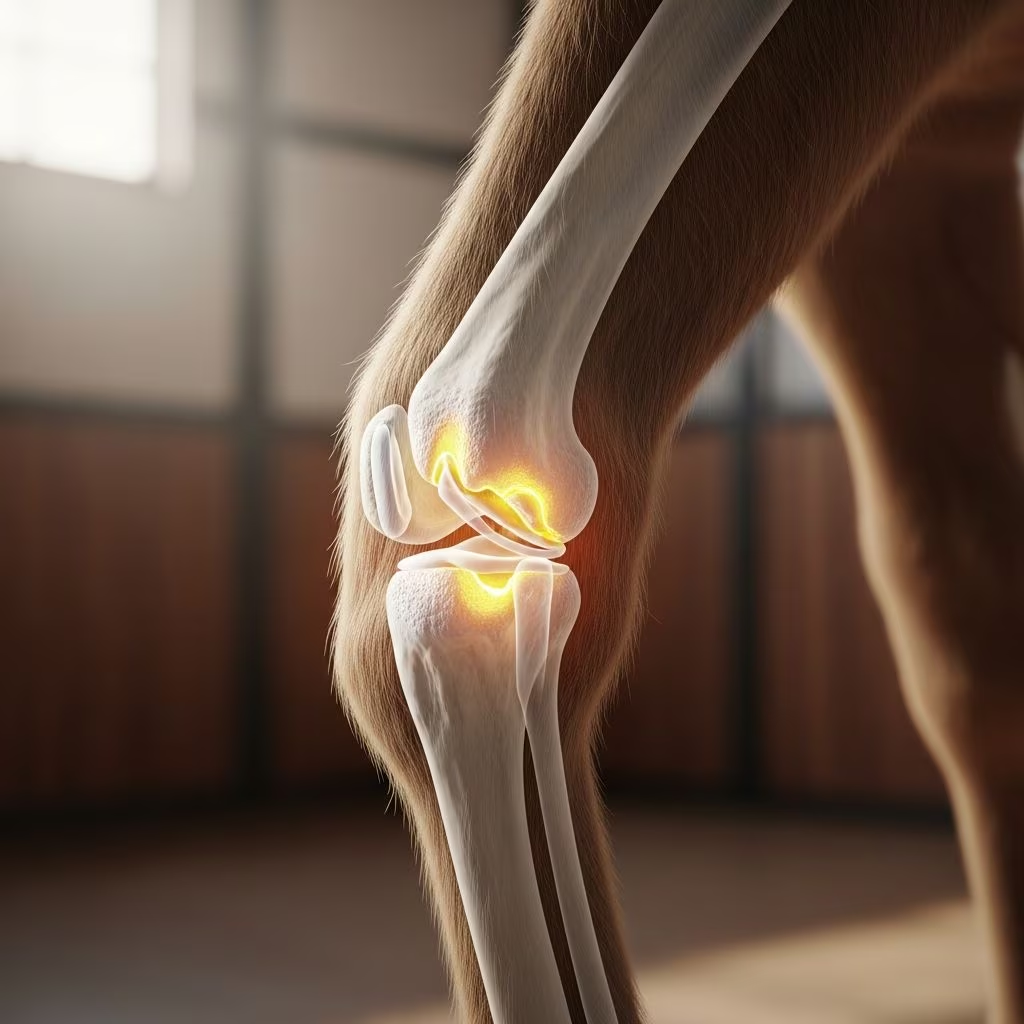
The Carpus and Its Critical Role in Equine Movement
The carpal joint complex represents one of the most important structures in equine locomotion, situated between the radius and metacarpal bones in the horse’s foreleg. This intricate anatomical region comprises three distinct articulations that work in concert to facilitate movement and absorb the tremendous forces generated during work and athletic activities. Understanding the structure and function of this joint system is essential for recognizing how degenerative changes develop and progress.
The carpal apparatus includes the antebrachiocarpal joint (formed between the distal radius and proximal row of carpal bones), the middle carpal joint (connecting the proximal and distal rows of carpal bones), and the carpometacarpal joint (linking the distal carpal row to the metacarpal bones). The antebrachiocarpal and middle carpal joints facilitate significant flexion and extension movements, while the carpometacarpal joint permits only minimal motion due to its structural design. Two distinct rows of cuboidal carpal bones are positioned between the radius and metacarpus, with each bone connected by specialized interosseous and transverse dorsal ligaments that provide structural stability.
The mechanical design of the carpal joint reflects millions of years of evolutionary optimization for weight-bearing and shock absorption. During loading, compression stress is absorbed through the intercarpal ligaments as carpal bones are separated by a wedge action, while extension is prevented by specialized stop facets located on the dorsal aspect of the carpal bones and supported by a stay apparatus. This sophisticated biomechanical system allows horses to handle repeated impact forces that exceed several times their body weight during movement at various gaits.
The Development of Degenerative Joint Disease in the Carpus
Osteoarthritis of the carpal joint represents a progressive degenerative condition characterized by deterioration of the articular cartilage, underlying bone, and surrounding soft tissues. Unlike acute injuries that develop suddenly, carpal osteoarthritis typically emerges over an extended period as cumulative damage to joint structures exceeds the body’s capacity for repair and remodeling. Multiple factors contribute to the initiation and progression of this disease, ranging from genetic predisposition and conformation abnormalities to training intensity and previous joint trauma.
The pathophysiological cascade of carpal osteoarthritis involves complex interactions between mechanical stress, inflammatory responses, and degenerative processes. Articular cartilage, which normally provides a smooth, frictionless surface for joint movement, gradually loses its protective qualities through fibrillation and erosion. As the cartilage layer thins and eventually exposes the underlying subchondral bone, the joint’s shock-absorbing capacity diminishes significantly. The body responds to this damage through production of new bone formation, which appears as osteophytes or bone spurs visible on radiographic examination.
Chronic synovitis, an inflammation of the synovial membrane that lines the joint capsule, frequently accompanies carpal osteoarthritis and perpetuates the degenerative process. Affected joints may demonstrate radiographic evidence of periosteal new bone formation on adjacent structures, particularly in cases where extensor carpi radialis synovitis occurs, manifesting as bone proliferation on the cranial distal aspect of the radius. This inflammatory response, while representing the body’s attempt at stabilization, actually contributes to further tissue damage and functional deterioration.
Clinical Presentation and Recognition of Carpal Disease
Horses affected by carpal osteoarthritis present with variable clinical signs depending on disease severity, duration, and which articular structures are primarily involved. The most common presentation includes lameness of the affected forelimb that may be subtle during initial stages but becomes increasingly apparent as disease progresses. Affected horses typically demonstrate shortened stride length, with some animals demonstrating obvious lameness at trot while appearing nearly normal at walk.
Visible swelling over the dorsal aspect of the carpus represents a hallmark sign of carpal joint disease. This swelling may be diffuse throughout the joint region or more localized to specific areas such as the tendon sheath, depending on which structures are involved. In acute inflammatory episodes, the swelling may appear relatively warm to palpation and respond with increased lameness to flexion of the joint. Chronic cases may develop thickened, fibrotic soft tissue that creates a permanent, cool swelling around the joint.
Pain response during physical examination provides important diagnostic information. Many affected horses demonstrate pain during forced flexion of the carpal joint, and some experience relief of lameness following application of local anesthesia to the joint region. The degree of pain often correlates with the inflammatory status of the joint rather than the absolute severity of radiographic changes, meaning that some horses with minimal radiographic evidence may experience considerable discomfort while others with extensive degenerative changes remain relatively functional.
Behavioral changes may accompany carpal osteoarthritis, including reluctance to stand on affected limbs, frequent shifting of weight during rest, and apparent stiffness that improves with exercise as synovial fluid lubricates the joint. Some horses demonstrate asymmetric weight distribution or adopt postural changes to minimize weight bearing on the affected carpus.
Diagnostic Approaches and Imaging Interpretation
Accurate diagnosis of carpal osteoarthritis requires integration of clinical examination findings with imaging evidence. Radiography remains the primary diagnostic imaging modality for carpal joint disease, allowing evaluation of both the joint surfaces and surrounding bone structures.
Radiographic Findings and Their Significance
Several radiographic projections provide complementary views necessary for thorough carpal evaluation. Standard lateral views demonstrate overall joint alignment and gross abnormalities, while dorsopalmar projections reveal medial-lateral relationships between carpal bones. Oblique views offer visualization of intercarpal articulations and help prevent misinterpretation of normal anatomical articulations as fractures—particularly important when distinguishing the articulation between the third and fourth carpal bones from lateral parasagittal fractures, or the articulation between the second and third carpal bones from medial parasagittal fractures.
Classical radiographic evidence of carpal osteoarthritis includes osteophyte formation along articular margins, subchondral bone sclerosis (increased bone density adjacent to the articular surface), and joint space narrowing. Soft tissue swelling visible on radiographs, particularly over the dorsal carpus, suggests active synovitis. Periosteal new bone formation on adjacent structures such as the radius provides evidence of chronic inflammatory changes.
Subchondral bone cysts represent another radiographic finding potentially associated with carpal joint disease. These appear as well-circumscribed circular or semicircular lucent (dark) areas within carpal bones, the distal radius, or proximal metacarpal bones, sometimes surrounded by an opacity. While some cystic lesions clearly communicate with the articular surface, others remain adjacent to the surface without obvious communication visible on radiographs. Secondary osteoarthritic changes may be evident surrounding these lesions.
Advanced Imaging Modalities
Ultrasound examination complements radiography by evaluating soft tissue structures including joint capsule thickness, synovial fluid characteristics, and tendon or ligament changes. Ultrasound proves particularly valuable for detecting subtle synovitis or soft tissue inflammation not always evident on radiographs.
Advanced imaging such as computed tomography (CT) or nuclear scintigraphy (bone scanning) may be employed in complex cases or when determining the functional impact of radiographic changes. Scintigraphy can identify increased metabolic activity in joints with active degeneration, sometimes revealing subclinical disease not yet apparent on radiographs.
Risk Factors and Predisposing Conditions
Multiple factors influence an individual horse’s susceptibility to developing carpal osteoarthritis:
- Conformation and Anatomy: Carpal conformation significantly influences joint soundness and longevity, with certain conformational variations predisposing to accelerated wear patterns
- Age and Use Intensity: Older horses and those subjected to intense athletic training accumulate greater cartilage damage over time
- Previous Trauma: Prior fractures, severe sprains, or other injuries to the carpal region substantially increase risk for subsequent osteoarthritis
- Genetic Predisposition: Certain bloodlines demonstrate higher incidence of carpal joint disease
- Training Methods: Excessive training intensity, inadequate conditioning periods, and improper loading progressions contribute to premature cartilage degeneration
- Surface and Footing: Work on excessively hard or uneven surfaces increases carpal joint stress
Management Strategies and Treatment Approaches
While osteoarthritis represents an irreversible degenerative process, multiple management strategies can slow disease progression, reduce pain, and maintain functional capability in affected horses.
Conservative Management Options
Rest and activity modification form the foundation of initial management, with the goal of reducing mechanical stress on affected joints while allowing inflammatory episodes to resolve. This may range from complete stall rest during acute flare-ups to controlled exercise that maintains muscle tone without exacerbating joint inflammation.
Weight management proves particularly important, as excessive body weight increases compressive forces through the carpal joint. Maintaining optimal body condition reduces stress on compromised cartilage and surrounding structures.
Farrier management including appropriate shoeing modifications can substantially impact carpal joint loading patterns. Adjustments to heel height, weight distribution, and breakover can reduce abnormal stresses through affected joints.
Pharmacological Interventions
Systemic anti-inflammatory medications including non-steroidal anti-inflammatory drugs (NSAIDs) reduce pain and inflammation, improving comfort and functional capability. Long-term NSAID use requires monitoring for gastrointestinal and renal side effects but remains a cornerstone of carpal osteoarthritis management.
Intra-articular therapeutic injections deliver medications directly into the affected joint, providing local anti-inflammatory effects while minimizing systemic exposure. Corticosteroid injections reduce synovitis and inflammatory pain, while hyaluronic acid products may improve synovial fluid viscosity and provide protective effects. Joint injections typically require professional administration under sterile conditions to prevent iatrogenic infection.
Systemic joint supplements containing glucosamine, chondroitin sulfate, or other constituents may provide modest protective effects, though evidence for efficacy varies considerably among products and individual horses.
Physical and Rehabilitative Approaches
Therapeutic exercise programs designed to maintain or rebuild muscle support around affected joints prove valuable. Controlled exercise helps preserve joint flexibility, maintains proprioceptive function, and reduces compensatory loading of other limbs. Swimming and treadmill exercise on forgiving surfaces allow exercise without excessive joint impact.
Physical therapies including cold laser, therapeutic ultrasound, and therapeutic massage may provide adjunctive benefits when incorporated into comprehensive management programs, though primary reliance should remain on evidence-based interventions.
Long-Term Prognosis and Functional Outcomes
The long-term prognosis for horses with carpal osteoarthritis depends on multiple factors including disease severity, structures involved, individual pain tolerance, intended use, and compliance with management recommendations. Many horses remain functionally sound for recreational and light work despite radiographic evidence of osteoarthritis, particularly when appropriate management strategies are consistently applied.
Progressive cases may eventually develop lameness severe enough to preclude ridden work, though some affected horses maintain acceptable comfort for ground handling and limited movement. The unpredictable nature of disease progression makes individual prognosis challenging to estimate, as some horses demonstrate long periods of stability while others experience rapid deterioration.
Early recognition and implementation of appropriate management during initial disease stages offers the best opportunity for preserving functional capability and maintaining quality of life. Owners should maintain realistic expectations regarding the irreversible nature of cartilage damage while recognizing that appropriate management can substantially prolong useful soundness.
Prevention Strategies and Risk Reduction
While genetic and conformational factors cannot be modified, several preventive approaches may reduce the risk or delay onset of carpal osteoarthritis:
- Gradual progression of training intensity with adequate recovery periods between work sessions
- Work on appropriate footing that provides adequate support without excessive concussion
- Maintenance of optimal body weight and physical fitness
- Regular farrier care with attention to proper hoof balance and shoeing
- Prompt treatment of injuries and inflammatory episodes to prevent chronic changes
- Periodic veterinary evaluation to detect early signs of joint disease
References
- The Equine Carpus — Veterian Key. Accessed February 2026. https://veteriankey.com/the-equine-carpus/
- Equine Forelimb Carpus Example 1 – Imaging Anatomy — University of Illinois College of Veterinary Medicine. Accessed February 2026. https://vetmed.illinois.edu/imaging_anatomy/equine/forelimb/carpus/ex01/carpus01-eq.html
- Carpal Joint Problems – Large Animal Surgery – Supplemental Notes — University of Minnesota Open Textbook Library. Accessed February 2026. https://open.lib.umn.edu/largeanimalsurgery/chapter/carpal-joint-problems/
- The Function of the Equine Carpal Joint: A Review — PubMed/National Center for Biotechnology Information. Accessed February 2026. https://pubmed.ncbi.nlm.nih.gov/16031805/
- Equine Carpal Conformation and Joint Soundness Variations — Open Veterinary Journal. Accessed February 2026. https://www.openveterinaryjournal.com/index.php?mno=144714&html=1
- The Carpus and Tarsus: Function and Construction — Horses Inside Out. Accessed February 2026. https://www.horsesinsideout.com/product-page/the-carpus-and-tarsus



