

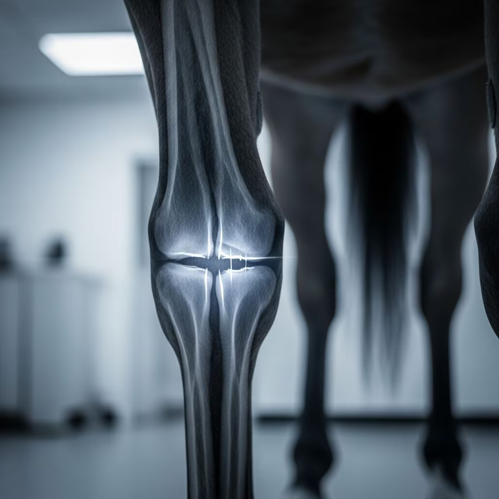
Fractures of the carpal bones represent a significant challenge in equine orthopedics, particularly among high-performance horses such as racehorses and jumpers. These injuries often arise from intense physical demands, leading to lameness that can sideline animals for months. Understanding the anatomy, mechanisms of injury, and management strategies is crucial for veterinarians and horse owners to facilitate recovery and prevent recurrence.
Understanding the Equine Carpus Anatomy
The carpus, or knee joint in horses, is a complex structure comprising three main rows of bones: the radial, intermediate, and accessory carpal bones proximally, followed by the ulnar, second, third, and fourth carpal bones distally. This arrangement allows for stability and flexibility during locomotion. The middle carpal joint and radiocarpal joint are primary sites for load-bearing, making them vulnerable to traumatic forces during high-speed activities or falls.
Key bones prone to fracture include the third carpal bone, radial carpal bone, and accessory carpal bone. The accessory carpal bone, positioned on the caudal aspect, supports tendon passage and is susceptible to specific shear forces. Knowledge of these anatomical features aids in pinpointing fracture locations and tailoring treatments.
Common Causes and Risk Factors
Trauma from high-speed exercise is the predominant cause of carpal fractures, especially in young racehorses whose skeletal systems are still maturing. Repetitive stress leads to microdamage accumulation, culminating in overt fractures. Chip fractures, involving small osteochondral fragments, frequently occur on the dorsal joint surfaces due to hyperextension or compressive impacts.
- High-impact activities: Racing, jumping, and steeplechasing expose the carpus to extreme flexion and extension.
- Direct trauma: Kicks, falls, or collisions with obstacles can produce slab or complete fractures.
- Predisposing conditions: Poor bone conditioning, nutritional deficiencies, or underlying joint degeneration increase susceptibility.
In thoroughbreds and quarter horses, genetic factors may contribute, with certain bloodlines showing higher incidence rates. Environmental factors like track surfaces also play a role; harder surfaces amplify impact forces.
Recognizing Clinical Signs
Horses with carpal fractures typically exhibit acute or progressive lameness, ranging from mild shortening of stride to non-weight-bearing. Swelling, heat, and joint effusion are hallmark signs, often accompanied by pain on flexion or direct palpation.
| Symptom | Description | Associated Fracture Type |
|---|---|---|
| Severe lameness | Sudden onset post-exercise, non-weight-bearing | Slab or complete fractures |
| Joint effusion | Fluid accumulation, visible distension | Chip fragments, accessory carpal |
| Pain on flexion | Resistance during carpal manipulation | All types, especially dorsal chips |
| Swelling/heat | Localized inflammation caudal or dorsal | Accessory carpal fractures |
Chronic cases may show subtle poor performance without overt lameness, masking the injury until advanced diagnostics are employed. Neurological signs like incoordination are rare but indicate severe displacement.
Diagnostic Approaches
Accurate diagnosis hinges on a combination of clinical examination and imaging. Initial assessment involves gait evaluation, flexion tests, and palpation to localize pain. Nerve blocks, such as ulnar blocks, help isolate the carpus.
Radiography remains the gold standard, revealing chip fragments, slab fractures, or displacement. Standard views include lateromedial, dorsopalmar, and flexed projections. Ultrasonography assesses soft tissue involvement, like tendon sheath effusion or fragments impinging on structures.
- X-rays: Identify fracture plane, fragment size, and joint involvement.
- Ultrasonography: Detects soft tissue changes and dynamic fragment movement.
- Advanced imaging: CT or MRI for complex cases, though less common in field settings.
Differentiating fractures from soft tissue injuries like desmitis requires serial imaging to monitor progression.
Types of Carpal Fractures
Osteochondral Chip Fractures
These small fragments from bone and cartilage are prevalent in racehorses, originating from the distal radial carpal or proximal third carpal bones. Trauma during fast exercise shears off dorsal projections, causing synovial irritation and lameness. Early intervention prevents osteoarthritis.
Slab Fractures
Extending fully across a bone, slab fractures most commonly affect the third carpal bone’s radial facet in frontal planes. They result from sagittal shear or compressive overload, leading to instability and rapid joint degeneration if untreated.
Accessory Carpal Bone Fractures
Less frequent but debilitating, these occur palmar to the lateral groove, often from falls on flexed limbs or the ‘nutcracker effect’ between radius and metacarpus. Seventeen of nineteen reported cases showed frontal plane fractures, with many achieving fibrous union despite non-bony healing. Lameness is severe, with carpal sheath effusion common.
Treatment Strategies
Management varies by fracture type and severity. Conservative approaches suit non-displaced chips or stable accessory fractures, while surgery is imperative for slabs.
Conservative Management:
- Stall rest: 6-12 months to promote fibrous union.
- Anti-inflammatories: NSAIDs reduce swelling and pain.
- Supportive therapy: Cold therapy, shockwave, or bisphosphonates enhance healing.
Surgical Interventions:
- Arthroscopic removal: For chips, fragments are debrided to restore joint congruity.
- Lag screw fixation: Stabilizes slab fractures.
- Tenoscopy: Addresses sheath complications in accessory fractures.
Post-operative care includes controlled exercise progression and monitoring for complications like infection or laminitis.
Prognosis and Long-Term Outcomes
Prognosis is favorable for chip fractures post-arthroscopy, with 70-90% of horses returning to racing. Slab fractures carry guarded outcomes due to osteoarthritis risk, though fixation improves success. Accessory fractures often heal via fibrous union, enabling soundness in 80% of cases, including competitive return.
Factors influencing recovery include age, fracture chronicity, and owner compliance. Regular radiographic follow-ups track healing.
Prevention Measures for Horse Owners
Proactive strategies mitigate risks:
- Conditioning programs: Gradual intensity buildup strengthens bones.
- Nutrition: Balanced calcium-phosphorus ratios and vitamin D support density.
- Farriery: Proper shoeing reduces uneven stress.
- Regular vet checks: Early detection of stress remodeling prevents progression.
Track management, like softer surfaces, further aids prevention.
Frequently Asked Questions (FAQs)
What is the most common carpal fracture in racehorses?
Osteochondral chip fractures on dorsal joint surfaces, caused by high-speed trauma.
How long does recovery take for accessory carpal fractures?
Typically 6-12 months with rest, leading to fibrous union and soundness.
Can horses return to racing after carpal slab fractures?
Yes, with surgical fixation, though osteoarthritis may limit full performance.
What are early signs of carpal issues?
Mild lameness, joint swelling, and pain on flexion; prompt imaging is key.
Is surgery always necessary?
No, stable fractures heal conservatively, but displaced ones require intervention.
References
- Disorders of the Carpus and Metacarpus in Horses — MSD Veterinary Manual. 2023. https://www.msdvetmanual.com/horse-owners/bone-joint-and-muscle-disorders-in-horses/disorders-of-the-carpus-and-metacarpus-in-horses
- Fracture of the Carpal Bones in Horses — Merck Veterinary Manual. 2023. https://www.merckvetmanual.com/musculoskeletal-system/disorders-of-the-carpus-in-horses/fracture-of-the-carpal-bones-in-horses
- Fractures in Horses — World Horse Welfare. 2023. https://www.worldhorsewelfare.org/advice/fractures-in-horses
- Fractures of the Accessory Carpal Bone in the Horse — Mad Barn Research Bank (PubMed). 1990-04-28. https://madbarn.com/research/fractures-of-the-accessory-carpal-bone-in-the-horse/
- Accessory Carpal: Fracture in Horses — Vetlexicon Equis. 2023. https://www.vetlexicon.com/equis/surgery-orthopedic/articles/accessory-carpal-fracture/



