

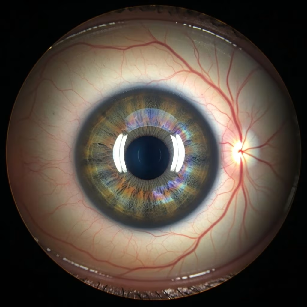
The ocular fundus, encompassing the retina, choroid, and optic disk, plays a crucial role in a dog’s vision. Disruptions in these structures can lead to partial or complete vision loss, making early recognition essential for pet owners. This article delves into key disorders affecting these areas, drawing from veterinary insights to help you identify signs and seek timely care.
Understanding the Canine Ocular Fundus
The retina is the light-sensitive layer at the back of the eye, converting light into neural signals for the brain. The choroid supplies blood and nutrients to the retina, while the optic disk serves as the exit point for optic nerve fibers. In dogs, these components appear distinct during fundoscopic exams: the retina shows a tapetal region with a reflective sheen in most breeds and a non-tapetal area that’s darker. Abnormalities here often manifest as vision decline, dilated pupils, or reluctance to navigate familiar spaces.
Breeds like Collies, Shelties, and Border Collies face heightened risks due to genetic predispositions, but any dog can develop acquired issues from trauma, infection, or systemic diseases like hypertension.
Progressive Retinal Atrophy: A Leading Genetic Threat
Progressive retinal atrophy (PRA) represents one of the most prevalent inherited retinal degenerations in dogs. This condition causes a gradual breakdown of photoreceptor cells, starting with rods responsible for night vision, followed by cones for daylight and color perception. Affected dogs initially struggle in dim light, bumping into objects at dusk, before progressing to total blindness over months or years. Remarkably, their eyes often appear normal externally, with no pain or inflammation.
PRA is documented across over 100 breeds, with early onset forms appearing before age three and late onset after five years. Genetic testing now enables breeders to screen carriers, reducing incidence. Diagnosis relies on electroretinography (ERG), which measures retinal electrical responses, alongside fundoscopy revealing thinned vessels and hyper-reflective retina.
- Common breeds affected: Labrador Retrievers, Australian Shepherds, Miniature Poodles
- Management: No cure exists; antioxidants like vitamin E may slow progression, but supportive care focuses on environmental adaptations.
Retinal Detachment and Its Urgent Implications
Retinal detachment occurs when the retina separates from underlying supportive tissues, often due to fluid accumulation under systemic conditions such as kidney disease, heart failure, or uncontrolled hypertension. Trauma from blunt force can also shear the retina. Dogs exhibit sudden blindness in one or both eyes, with pupils that remain dilated even in light and may show hemorrhage or folds on exam.
Treatment targets the underlying cause: blood pressure medications for hypertension or diuretics for fluid overload. Surgical reattachment is rare in veterinary practice due to complexity and cost, but partial recovery is possible if addressed promptly. Prognosis hinges on rapid intervention; delays often result in permanent vision loss.
| Detachment Cause | Symptoms | Treatment Approach |
|---|---|---|
| Hypertension | Sudden bilateral blindness | Antihypertensives |
| Trauma | Unilateral vision loss, hemorrhage | Surgical evaluation |
| Systemic disease | Variable onset | Address primary illness |
Choroidal Disorders: From Hypoplasia to Degeneration
The choroid’s pigmentation and vascular integrity are vital for retinal health. Choroidal hypoplasia, part of Collie eye anomaly (CEA), features localized thinning or absence of choroidal tissue, appearing as pale fundus patches. This congenital defect, inherited recessively, correlates with retinal detachment risk in 20-30% of cases, typically by two years of age. CEA primarily strikes herding breeds, where DNA tests confirm carrier status for breeding decisions.
Choroidal degeneration arises secondarily from long-term retinal diseases or inflammation, leading to vessel atrophy and pigment clumping. Unlike hypoplasia, it’s acquired and non-heritable.
- Key signs: Tapetal hyper-reflectivity, pigment migration
- Breeds at risk: Rough/Smooth Collies, Australian Shepherds
Optic Disk Abnormalities and Neuropathy
The optic disk, or optic nerve head, transmits visual signals. Optic neuritis involves inflammation from infections, immune-mediated diseases, or toxins, causing acute blindness, mydriasis, and a swollen, hyperemic disk on exam. Prognosis improves with steroids and addressing causes like fungal infections in endemic areas.
Optic nerve hypoplasia is a congenital malformation with a small or absent disk, leading to blindness from birth. Colobomas present as excavated pits in the disk, potentially enlarging painfully and risking detachment. These developmental issues cluster in breeds like Labrador Retrievers and Miniature Poodles.
Atrophy develops post-trauma or ischemia, shrinking the disk and vessels, confirming with ERG or visual evoked potentials.
Diagnostic Tools for Fundus Evaluation
Veterinarians employ indirect ophthalmoscopy with a 28D lens for panoramic fundus views, revealing subtle changes missed by direct methods. Fluorescein angiography highlights vascular leaks, while optical coherence tomography (OCT) provides cross-sectional retinal layers in advanced clinics. Tonometry measures intraocular pressure to rule out glaucoma overlaps, and B-scan ultrasound assesses detachments when media opacity hinders visualization.
Treatment Strategies and Prognosis
Therapy varies by disorder: PRA management is palliative, emphasizing quality of life through scent training and stable home layouts. Detachments demand systemic workups, including blood pressure checks and urinalysis. Inflammatory conditions respond to topical or systemic steroids, immunosuppressants like cyclosporine, or antibiotics for infections.
Surgical interventions, such as cryotherapy for peripheral detachments or vitrectomy for select cases, are specialist-driven. Regular monitoring prevents progression in hypoplasia carriers. Overall, early detection via annual exams in at-risk breeds markedly improves outcomes.
Prevention Through Breeding and Routine Care
Genetic screening via panels from labs like Optigen prevents PRA and CEA propagation. Avoid breeding affected dogs, per guidelines from the Orthopedic Foundation for Animals (OFA). Daily eye hygiene—gentle saline wipes—reduces irritation risks. Protect active dogs with goggles during rough play, and control systemic diseases via vet check-ups.
Puppy wellness visits should include pupillary light reflexes and menace response tests for early anomalies.
Frequently Asked Questions (FAQs)
Can retinal disorders be cured in dogs?
Most retinal degenerations like PRA are incurable but manageable; detachments may partially resolve with prompt treatment of underlying causes.
What breeds are prone to choroidal hypoplasia?
Primarily Collie-related breeds, Shetland Sheepdogs, and some herding types; genetic testing is recommended.
How do I know if my dog has optic nerve issues?
Signs include sudden blindness, absent pupillary response; a veterinary ophthalmologist confirms via fundoscopy and ERG.
Is surgery common for fundus disorders?
Rarely, due to technical challenges; medical management predominates.
Can older dogs develop these problems?
Yes, atrophy and degeneration increase with age, alongside systemic factors.
Key Takeaways for Pet Owners
- Monitor for vision changes like hesitation in dark or collisions.
- Schedule breed-specific eye screenings annually.
- Seek immediate vet care for acute blindness or eye pain.
- Support breeding programs with certified clear genetics.
References
- Disorders of the Retina, Choroid, and Optic Disk (Ocular Fundus) in Dogs — Merck Veterinary Manual. 2023. https://www.merckvetmanual.com/dog-owners/eye-disorders-of-dogs/disorders-of-the-retina-choroid-and-optic-disk-ocular-fundus-in-dogs
- 22 Common Dog Eye Problems — PetMD. 2024-02-15. https://www.petmd.com/dog/general-health/evr_dg_eye_problems_in_dogs
- A Guide to Common Eye Problems — Calder Vets. 2023. https://www.caldervets.co.uk/pet-help-advice/dogs/eye-care-for-dogs/55-a-guide-to-common-eye-problems
- Canine Eye Health — AKC Canine Health Foundation. 2022-06-01. https://www.akcchf.org/disease-history/canine-eye-health/
- Hereditary Eye Disease in Dogs — British Veterinary Association. 2019-07-10. https://www.bva.co.uk/media/2811/20190710-chs-eye-disease-guide-0719-v1-web.pdf



