

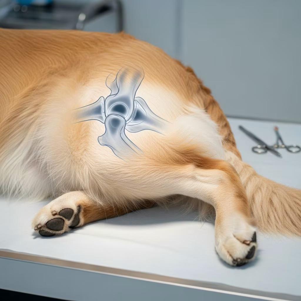
Hip luxation in dogs, where the femoral head slips out of the acetabulum socket, demands prompt attention to prevent lasting damage and pain. This orthopedic issue, often stemming from trauma, affects mobility but responds well to timely veterinary care.
Understanding the Canine Hip Joint Anatomy
The hip joint forms a ball-and-socket structure, with the femur’s rounded head fitting into the pelvis’s acetabulum for fluid movement. Ligaments and muscles stabilize this setup, enabling running, jumping, and standing. Disruption occurs when forceful events eject the femoral head, leading to luxation.
- Key Components: Femoral head (ball), acetabulum (socket), joint capsule, and supporting ligaments.
- Luxation types include cranial (forward), ventral (downward), and caudal (backward), each influencing treatment choices.
Pre-existing weaknesses like shallow sockets from hip dysplasia heighten vulnerability, though trauma remains the primary trigger.
Primary Causes of Hip Luxation
Trauma accounts for most cases, involving high-impact events that overwhelm joint stability. Dogs hit by vehicles, falling from elevations, or colliding during play face elevated risks.
| Cause Type | Examples | Risk Factors |
|---|---|---|
| Traumatic | Car accidents, falls, fights | Active/outdoor dogs |
| Congenital | Hip dysplasia, lax ligaments | Large breeds (e.g., Labs, German Shepherds) |
| Degenerative | Arthritis erosion | Aging or chronic joint issues |
Unlike developmental dysplasia, luxation typically presents acutely post-injury, causing immediate distress.
Recognizing Symptoms in Your Dog
Owners often spot hindlimb lameness first, where the dog refuses weight-bearing or hobbles with toe-touching gait. The leg may appear shortened, externally rotated, or flexed upward.
- Acute Signs: Severe pain on touch, reluctance to rise, yelping during movement.
- Chronic Indicators: Muscle wasting, subtle limp, abnormal posture.
- Behavioral shifts like withdrawal or aggression when leg is approached signal discomfort.
Small breeds might mask symptoms initially, but any gait change warrants examination. Delays risk avascular necrosis or arthritis.
Diagnostic Process for Confirmation
Veterinarians start with physical exams, testing joint stability under sedation due to pain. Radiographs reveal dislocation direction, fractures, or soft tissue damage.
- History review: Recent trauma?
- Palpation: Assess range, crepitus.
- Imaging: X-rays pre- and post-manipulation.
- Advanced: CT/MRI for complexities.
Fractures complicate prognosis, prioritizing stabilization first.
Non-Surgical Treatment Options
Closed reduction manually repositions the joint under anesthesia, followed by immobilization. Success hinges on early intervention.
- Ehmer Sling: Bandage folds leg to maintain position; weekly X-rays verify stability.
- Pain control: NSAIDs, opioids.
- Strict crate rest: 2-4 weeks minimal activity.
Viable for recent, uncomplicated cases without fractures; re-luxation risk drops post-14 days if stable.
Surgical Interventions Explained
When non-surgical fails or damage is severe, surgery restores function. Options scale by case severity.
| Procedure | Description | Best For | Recovery Time |
|---|---|---|---|
| Femoral Head Ostectomy (FHO) | Removes femoral head, forms false joint via scar tissue. | Small dogs, cost-effective salvage. | 4-6 weeks |
| Total Hip Replacement (THR) | Prosthetic joint installation. | Young/large dogs, severe damage. | 8-12 weeks |
| Toggle Pin/Capsulorrhaphy | Repairs ligaments, adds stabilizing pin. | Acute, stable reductions. | 6-8 weeks |
FHO yields near-normal function; THR offers longevity but higher cost. Post-op, arthritis may develop mildly.
Step-by-Step Recovery and Rehabilitation
Post-treatment, controlled activity prevents re-injury. Pain meds and rest dominate early phases.
- Weeks 1-2: Crate confinement, sling if used.
- Weeks 3-6: Leashed walks, passive range exercises.
- Ongoing: Physical therapy, weight management.
Hydrotherapy builds strength without stress. Monitor for swelling or instability; full activity resumes at 3 months typically.
Preventive Strategies for Hip Health
Minimize risks through breed-aware care. Large breeds benefit from dysplasia screening, joint supplements.
- Supervised play, avoid heights.
- Maintain ideal weight to reduce joint load.
- Regular vet checks for at-risk dogs.
Early dysplasia management via diet, exercise curbs luxation odds.
Prognosis and Long-Term Outlook
With swift treatment, most dogs regain 90-100% function. Untreated cases lead to chronic lameness, DJD. Surgical success exceeds 90% for FHO/THR.
Frequently Asked Questions
Can a dog’s hip fix itself?
No, luxation requires intervention; unmanaged cases form painful false joints.
How much does hip surgery cost for dogs?
Varies by procedure: FHO $1,500-$4,000; THR $5,000-$7,000, excluding rehab.
Which dogs are prone to hip luxation?
Large breeds with dysplasia like Retrievers, Shepherds; trauma affects all.
What if my dog limps after treatment?
Contact vet; may indicate re-luxation or infection needing prompt imaging.
Is physical therapy essential post-surgery?
Highly recommended to optimize mobility and prevent atrophy.
Supporting Your Dog Through Recovery
Emotional support aids healing; ramps, orthopedic beds ease strain. Track progress with photos, consult specialists for complexities.
References
- Dog Hip Dislocation – A Complete Guide — Zoomadog. 2023. https://zoomadog.co.uk/blogs/blog/dog-hip-dislocation-a-guide
- Hip Dislocation In Dogs: Everything You Need To Know — Kingsdale Animal Hospital. 2024-10-15. https://www.kingsdale.com/hip-dislocation-in-dogs-everything-you-need-to-know
- Dislocated Hips in a Dog: From causes to Correction — Long Beach Animal Hospital. 2023. https://lbah.com/canine/hip-dislocation/
- Hip Dislocation and Post-Operative Care in Dogs — VCA Animal Hospitals. 2025-01-10. https://vcahospitals.com/know-your-pet/hip-dislocation-and-post-operative-care-in-dogs
- Hip Luxation — American College of Veterinary Surgeons. 2024. https://www.acvs.org/small-animal/hip-luxation/



