

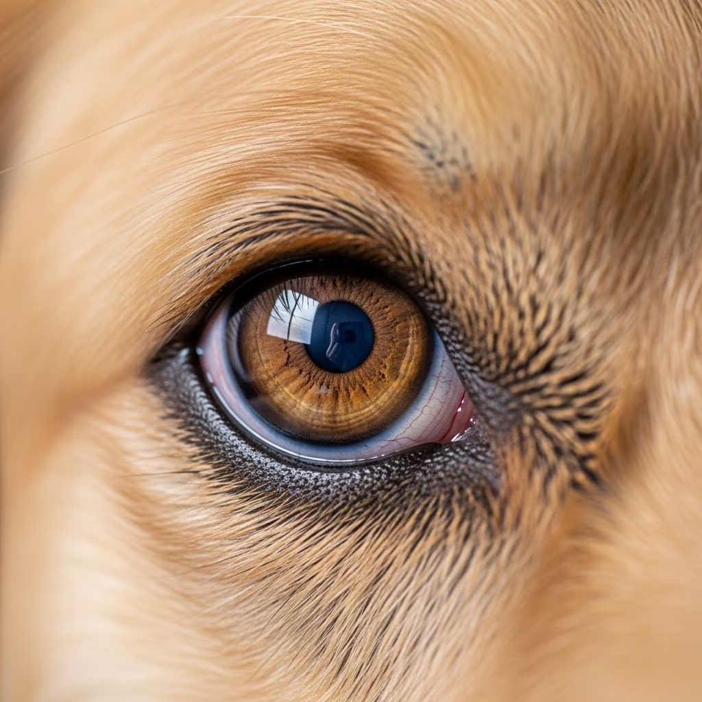
Glaucoma represents one of the most serious eye conditions affecting dogs, yet many owners remain unaware of its subtle warning signs until significant vision loss has already occurred. This condition develops when fluid within the eye fails to drain properly, resulting in elevated intraocular pressure that gradually damages the retina and optic nerve. Unlike some eye conditions that announce themselves dramatically, glaucoma often progresses silently, making early detection and intervention critical for preserving your dog’s sight.
What Happens Inside Your Dog’s Eye
To understand glaucoma, it helps to know how the eye normally functions. The eye continuously produces a clear fluid called aqueous humor that maintains the eye’s shape and supplies nutrients to internal structures. In a healthy eye, this fluid drains through microscopic channels at the base of the iris, maintaining a delicate pressure balance. Normal intraocular pressure ranges from 10 to 20 millimeters of mercury (mmHg). When drainage becomes impaired, pressure accumulates, and sustained elevation above 25 mmHg may indicate glaucoma, while readings exceeding 35 mmHg typically cause significant discomfort.
The consequences of elevated pressure are profound. The optic nerve, responsible for transmitting visual information to the brain, cannot withstand prolonged pressure elevation. Progressive nerve fiber death leads to irreversible vision loss, and without treatment, complete blindness can develop. The timeline varies considerably—some dogs experience rapid vision deterioration within days or weeks, while others face a slower, chronic progression over months or years.
Primary Glaucoma: The Genetic Factor
Primary glaucoma stems from inherited genetic mutations affecting the eye’s drainage system. Dogs born with this condition have structural abnormalities in the drainage area, a condition called goniodysgenesis, characterized by narrow or completely closed drainage ducts. These anatomical defects are present from birth but may not cause problems until the dog reaches middle age or beyond.
Certain breeds face substantially elevated risk for developing primary glaucoma. High-risk breeds include Cocker Spaniels, Basset Hounds, Beagles, Siberian Huskies, Dalmatians, Chow Chows, and Samoyed dogs. If your dog belongs to one of these breeds, vigilant monitoring becomes especially important. Primary glaucoma can affect one or both eyes, though it often begins in one eye before potentially developing in the other.
The genetic nature of primary glaucoma means that once one eye develops the condition, the other eye faces significant risk. Many veterinarians begin preventive eyedrop treatment in the unaffected eye to delay pressure elevation and catch any developing problems at their earliest stages.
Secondary Glaucoma: Underlying Causes
Secondary glaucoma develops as a consequence of another eye disease or injury and represents the most common form of glaucoma in dogs. Unlike primary glaucoma, secondary glaucoma can affect any dog, regardless of breed or genetic predisposition. This type typically presents more acutely and is more likely to be noticed by observant owners because the sudden onset often produces more noticeable symptoms.
Several underlying conditions can trigger secondary glaucoma:
- Lens-related problems: When cataracts develop, they can leak proteins into the eye, triggering inflammation that damages drainage pathways. Lens luxation—where the lens slips out of its normal position—can physically block fluid drainage, causing rapid pressure elevation.
- Inflammation: Uveitis, inflammation of the eye’s interior structures, can cause scarring and permanent blockage of drainage ducts. Inflammatory cells accumulate and damage the delicate drainage system.
- Eye injuries: Trauma can trigger swelling, bleeding, and inflammation that disrupts the normal fluid balance within the eye.
- Eye tumors: Masses inside the eye obstruct normal fluid circulation and can lead to rapidly elevated pressure.
- Systemic infections: Certain fungal diseases can cause eye inflammation and secondary glaucoma alongside systemic illness.
Secondary glaucoma typically affects only one eye, though addressing the underlying cause may prevent similar problems in the other eye.
Recognizing the Warning Signs
The challenge with canine glaucoma lies in its often-subtle presentation. Dogs may adapt remarkably well to chronic discomfort, masking symptoms that should trigger concern. Early detection requires understanding both the obvious and more subtle warning signs.
| Sign Category | Observable Symptoms |
|---|---|
| Physical Eye Changes | Cloudiness or bluish discoloration of the cornea, excessive redness, bloodshot appearance, bulging or swollen eye, enlarged or dilated pupil |
| Behavioral Changes | Decreased appetite, reduced activity level, lethargy, reluctance to play or move normally |
| Pain Indicators | Excessive squinting, eye discharge or tearing, sensitivity to light, pawing at the eye or face |
| Vision Loss Signs | Hesitation when moving through dark areas, bumping into objects, decreased response to thrown toys or treats, reluctance to navigate new environments |
Symptoms may develop gradually or appear suddenly. Dogs with acute glaucoma may show pain-related signs like squinting, excessive tearing, and behavioral changes within days. Chronic cases may progress so slowly that owners don’t notice gradual vision loss until the affected eye has already sustained significant damage. In some cases, owners only become aware of glaucoma after their dog has lost sight in one eye and vision problems appear in the other eye.
The Diagnostic Process
Accurate diagnosis requires more than visual inspection. Your veterinarian will perform several tests to confirm glaucoma and identify any underlying causes. The primary diagnostic tool is tonometry, which measures intraocular pressure using a specialized instrument. This painless test takes just seconds and provides critical pressure readings that confirm whether elevation has occurred.
Beyond pressure measurement, your veterinarian may recommend:
- Gonioscopy: This examination directly visualizes the drainage angle using a specialized lens to assess whether the drainage structures appear normal, narrow, or closed—particularly important for identifying primary glaucoma.
- Ophthalmoscopy: A detailed examination of the retina and optic nerve to determine whether damage has occurred and assess the severity of any nerve fiber loss.
- Ultrasound imaging: When the eye is too cloudy for direct examination, specialized ultrasound can detect lens displacement, tumors, retinal detachment, or bleeding that might explain secondary glaucoma.
- Fungal and bacterial testing: If systemic infection is suspected as the underlying cause, appropriate testing may be performed.
If your veterinarian suspects primary glaucoma in one eye, gonioscopy of the other eye can provide valuable information about its future risk, helping guide preventive strategies.
Medical Management Approaches
Initial glaucoma treatment focuses on rapidly reducing intraocular pressure through medications. The goal is to prevent further optic nerve damage and preserve remaining vision. Several medication classes work through different mechanisms:
Prostaglandin analogue drops (such as latanoprost, travoprost, and bimatoprost) represent the most effective medications for rapidly lowering pressure in emergency situations. These drugs increase fluid drainage from the eye and can bring dangerously elevated pressures down to safer levels relatively quickly.
Carbonic anhydrase inhibitors (such as dorzolamide and brinzolamide) decrease the amount of fluid the eye produces, reducing overall pressure. These are often considered the preferred long-term treatment option and may be administered as eye drops or, in some cases, as oral medications.
Beta-blocker drops (such as timolol) slow fluid production and improve drainage, contributing to overall pressure reduction.
Many dogs require multiple medications administered several times daily to maintain adequate pressure control. Your veterinarian will develop a customized regimen based on your dog’s specific response and needs. Importantly, these medications manage glaucoma—they don’t cure it. As the condition tends to progress over time, the effectiveness of medication may decline, necessitating adjustments or additional therapies.
Pain management is a crucial but sometimes overlooked component of treatment. Glaucoma is painful, and your veterinarian will prescribe appropriate painkillers alongside pressure-reducing medications to keep your dog comfortable.
When Surgery Becomes Necessary
When medical management fails to control pressure adequately, or when the medication schedule becomes impractical, surgery may be recommended. A veterinary ophthalmologist specializes in glaucoma surgical procedures and can explain which options might benefit your dog.
Vision-sparing surgeries aim to preserve or maintain vision in the affected eye:
- Laser cyclodestruction (endocyclophotocoagulation) destroys the cells that produce aqueous humor, reducing fluid production significantly.
- Shunt or implant surgery creates alternative drainage pathways, allowing fluid to escape the eye and reducing pressure.
- Micropulse laser and micro-invasive glaucoma surgery (MIGS) represent newer techniques being adapted for canine use from human ophthalmology.
Comfort-promoting surgeries relieve pain in eyes that have already lost vision or when other interventions have failed. These include prosthetic eye implants or injection of medications that permanently reduce fluid production. In severe cases where pain cannot be adequately managed, removal of the affected eye (enucleation) may be recommended. While this sounds drastic, dogs adapt remarkably well to single-eye vision, and the procedure eliminates chronic pain.
Surgical success rates vary, and your veterinary ophthalmologist will discuss realistic expectations for your dog’s specific situation.
The Critical Importance of Monitoring
Once glaucoma is diagnosed and treatment begins, consistent follow-up monitoring becomes essential. Pressure readings can change without obvious external signs, and permanent vision loss may develop if pressure remains elevated for extended periods. Initially, your veterinarian will recommend frequent follow-up examinations—sometimes weekly or every few weeks—to ensure your dog is responding adequately to treatment and to identify any need for medication adjustments.
As treatment stabilizes, the frequency of monitoring visits may decrease, but regular check-ups remain crucial indefinitely. These appointments allow your veterinarian to catch any pressure increases early and maintain vision as long as possible. Some dogs will eventually require surgical intervention despite medical management, and regular monitoring helps identify the optimal timing for these decisions.
When Glaucoma Becomes an Emergency
Certain situations demand immediate emergency veterinary care. Contact an emergency veterinarian immediately if you notice a suddenly bulging eye, acute onset of blindness, intense redness, or signs of severe pain including severe squinting or unresponsiveness. Eye pressures above 40–50 mmHg require emergency treatment to prevent rapid, irreversible vision loss. Acute glaucoma can progress from symptom onset to complete blindness within hours, making rapid intervention critical.
Preventive Strategies for High-Risk Dogs
For dogs belonging to breeds at higher risk for primary glaucoma, preventive measures can make a meaningful difference. Annual or biannual eye examinations by your veterinarian or an ophthalmologist allow early detection of pressure elevation before vision loss occurs. If your dog develops glaucoma in one eye, immediately beginning pressure-reducing eyedrops in the other eye—even if it appears healthy—can delay the onset of glaucoma and facilitate early detection if it does develop.
Maintaining overall eye health reduces secondary glaucoma risk. Promptly addressing eye injuries, treating inflammatory conditions, and managing systemic diseases that could affect eye health all contribute to glaucoma prevention.
Living with a Dog That Has Glaucoma
A glaucoma diagnosis is not a death sentence for your dog’s quality of life. With appropriate treatment and monitoring, many dogs maintain useful vision for extended periods. Some eventually experience vision loss despite optimal treatment, but dogs adapt remarkably well to blindness, relying on their other senses and their established knowledge of their environment.
Commitment to the treatment regimen your veterinarian recommends—whether medication administration, frequent monitoring visits, or eventual surgical intervention—significantly influences outcomes. Each dog’s response to treatment is individual, and your veterinarian will work with you to optimize your dog’s comfort and vision preservation.
References
- Glaucoma in Dogs: What Is It, and What Are the Symptoms? — PetMD. 2024. https://www.petmd.com/dog/conditions/eyes/glaucoma-dogs-what-it-and-what-are-symptoms
- Glaucoma — Cornell University College of Veterinary Medicine, Riney Canine Health Center. 2024. https://www.vet.cornell.edu/departments-centers-and-institutes/riney-canine-health-center/canine-health-topics/glaucoma
- Glaucoma in Dogs: Causes, Symptoms and Treatment Options — CareCredit Well-U. 2024. https://www.carecredit.com/well-u/pet-care/dog-glaucoma/
- Canine Glaucoma — Vision for Animals. 2024. https://www.visionforanimals.org/canine-glaucoma/
- Glaucoma in Dogs — VCA Animal Hospitals. 2024. https://vcahospitals.com/know-your-pet/glaucoma-in-dogs
- Dog Eye Glaucoma: Causes, Symptoms & Surgery — Animal Eye Guys. 2022. https://www.animaleyeguys.com/site/blog/2022/11/15/dog-eye-glaucoma



