

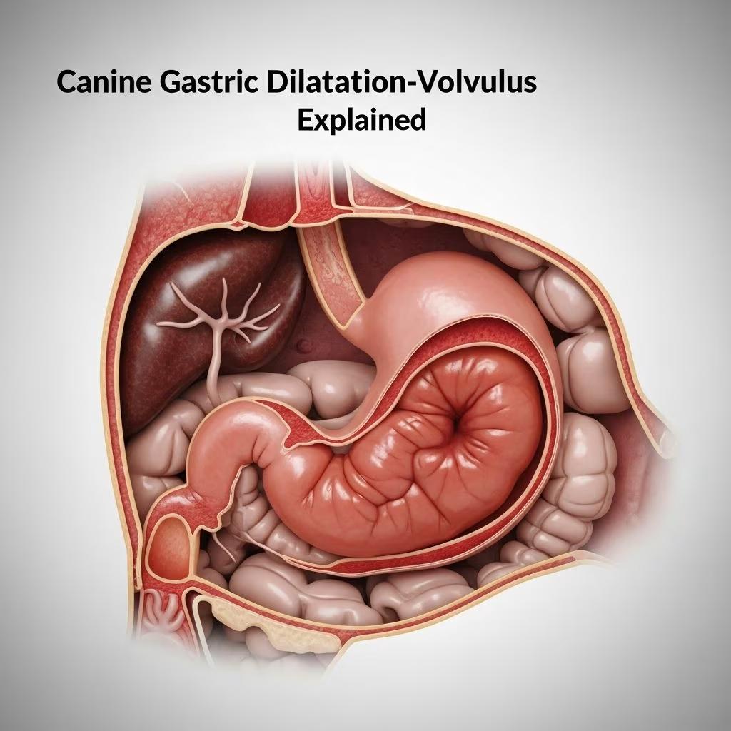
The canine digestive system relies on a simple, glandular stomach positioned in the abdominal cavity to process food efficiently. However, this organ can become susceptible to a severe emergency known as gastric dilatation-volvulus (GDV), where the stomach distends with gas and rotates on its axis, compromising blood flow and organ function. GDV demands immediate veterinary intervention to save the dog’s life.
Understanding the Structure of a Dog’s Stomach
Dogs possess a monogastric stomach, distinct from the multi-chambered systems in ruminants, serving as a reservoir for ingesta between the esophagus and small intestine. This organ features three primary regions: the cardia, where the esophagus enters; the fundus, forming the main body; and the pylorus, the exit to the duodenum.
The stomach’s position starts more on the left side of the abdomen, secured by peritoneal attachments like the gastrosplenic ligament and greater omentum, allowing flexibility but also mobility. Its muscular layers, particularly the thick circular layer along the greater curvature, enable grinding and mixing of food through peristaltic waves and segmentation. During digestion, the fundus relaxes to store food while the antrum churns contents, propelling small particles into the duodenum while retaining larger ones.
- Cardia region: Entry point for esophageal contents, featuring a sphincter to prevent reflux.
- Fundus and body: Dilatable areas for storage and initial microbial activity.
- Pylorus and antrum: Active grinding zones with sphincters controlling outflow.
Histologically, the glandular mucosa produces hydrochloric acid, pepsin, and mucus via tubular glands opening into gastric pits, preparing digesta for intestinal absorption. This design supports carnivorous diets but heightens vulnerability to distension.
How GDV Develops: The Mechanism of Stomach Distension and Twisting
GDV unfolds in stages: first, gastric dilatation occurs as the stomach fills with gas, fluids, and undigested food, often from swallowed air during rapid eating or exercise. Normal motility fails, trapping contents and causing progressive expansion.
The critical phase is volvulus, where the overfilled stomach rotates 90 to 360 degrees around its mesentery, typically clockwise. This twist occludes blood vessels, leading to tissue ischemia, shock, and potential rupture. Factors like the stomach’s left-sided starting position and attachments contribute to this rotation.
| Stage | Description | Consequences |
|---|---|---|
| Simple Dilatation | Gas buildup without twist | Discomfort, unproductive retching |
| Volvulus | Rotation and vascular compromise | Shock, necrosis, high mortality |
Motility patterns exacerbate the issue: fundus relaxation slows emptying, while antral contractions force contents against a closed pylorus, worsening distension.
Breeds and Factors Increasing GDV Susceptibility
Large, deep-chested breeds like Great Danes, German Shepherds, and Standard Poodles face elevated risks due to anatomical predisposition for stomach mobility. Age over seven years, stress, single large meals, and exercise post-feeding amplify chances.
- Genetic predisposition in giant breeds.
- Behavioral traits like gulping food.
- Environmental triggers such as high elevation or heat.
Males may be slightly more affected, and first-degree relatives of GDV cases show higher incidence, suggesting heritability.
Recognizing GDV Symptoms: From Mild Distress to Crisis
Early signs mimic indigestion: restless pacing, drooling, and futile vomiting attempts. Progression brings a distended abdomen, pale gums, rapid breathing, and collapse as shock sets in. Owners often note a sudden, unproductive retch, distinguishing it from typical vomiting.
Without treatment, symptoms escalate to weakness, arrhythmias from vagal stimulation, and death within hours. Differentiate from other issues like pancreatitis via the hard, tympanic belly on palpation.
Diagnostic Approaches in Veterinary Practice
Vets confirm GDV through physical exams revealing a bloated, painful abdomen and vital sign instability. Imaging is key: right lateral radiographs show a ‘double bubble’—gas in fundus and pylorus—diagnostic for volvulus. Ultrasound detects twists, while bloodwork reveals elevated lactate, acidosis, and electrolyte shifts signaling severity.
ECG monitors cardiac effects from splenic enlargement or electrolyte imbalance.
Emergency Stabilization and Surgical Intervention
Immediate action involves IV fluids, pain relief, and gastric decompression via orogastric tube or trocharization to release gas. Surgery (gastropexy) is essential: laparotomy derotates the stomach, assesses viability (non-viable tissue is resected), and anchors the stomach to the abdominal wall via permanent gastropexy to prevent recurrence.
Concurrent splenectomy occurs if the spleen is compromised. Postoperative care includes antibiotics, antiemetics, and monitoring for peritonitis or arrhythmias.
Postoperative Recovery and Long-Term Management
Hospital stays last 2-5 days with gradual feeding resumption using small, frequent meals. Complications like gastric necrosis (10-20% mortality) or re-dilatation (without gastropexy) require vigilance. Most dogs resume normal activity in weeks, with gastropexy reducing recurrence to under 5%.
Proactive Prevention Strategies for Dog Owners
Minimize risks by feeding multiple small meals from low bowls, avoiding exercise around mealtimes, and using slow-feeder bowls. Prophylactic gastropexy during spay/neuter is increasingly recommended for at-risk breeds.
- Split daily ration into 3-4 meals.
- Discourage gulping with puzzle feeders.
- Regular vet check-ups for early detection.
Frequently Asked Questions About GDV
What triggers GDV in dogs?
Common triggers include aerophagia from fast eating, large meals, genetic factors, and stress, leading to gas accumulation and rotation.
Can GDV resolve without surgery?
Simple bloat may decompress medically, but volvulus requires surgery; delays prove fatal.
Is gastropexy safe for all dogs?
It’s highly effective for deep-chested breeds, often performed preventively with minimal complications.
How quickly must I act on symptoms?
Seek emergency care immediately upon seeing distension and retching—survival drops after 2-6 hours.
Does diet influence GDV risk?
High-volume kibble or soy-based foods may contribute; opt for quality, measured portions.
Advances in GDV Research and Future Outlook
Ongoing studies explore genetic markers for screening and motility-modifying drugs. Enhanced diagnostics like point-of-care lactate testing improve outcomes. Owners armed with knowledge can protect their pets from this peril.
References
- Anatomy of the Canine Stomach, Small and Large Intestine — Veterinary Anatomy, YouTube. 2021-03-20. https://www.youtube.com/watch?v=8B3jdWmvAmU
- Stomach — Veterian Key. N/A. https://veteriankey.com/stomach-2/
- Anatomy of the Canine Digestive System — EasyAnatomy. N/A. https://easy-anatomy.com/canine-digestive-system/
- The Digestive System in Animals — Merck Veterinary Manual. N/A. https://www.merckvetmanual.com/digestive-system/digestive-system-introduction/the-digestive-system-in-animals
- Stomach – Veterinary Histology — Ohio State Pressbooks. N/A. https://ohiostate.pressbooks.pub/vethisto/chapter/8-stomach/



