

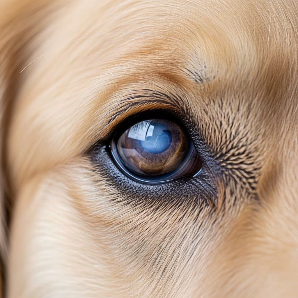
Vision is one of the most precious senses your dog relies on to navigate the world, interact with their environment, and maintain quality of life. When cloudiness develops in the lens—a condition known as cataracts—it can significantly impact how your canine companion experiences their surroundings. Understanding this common eye condition, its underlying causes, and available interventions is essential for any responsible dog owner seeking to maintain their pet’s ocular health and overall wellbeing.
What Are Cataracts and How Do They Develop?
A cataract occurs when the eye’s lens becomes opaque or cloudy, preventing light from passing through to the retina. The lens is normally clear and transparent, allowing light rays to focus properly on the retinal tissue at the back of the eye. This cloudiness develops due to alterations in either the water balance within the lens or changes to the protein structures that compose lens tissue. When light cannot reach the retina effectively, dogs experience diminished vision that resembles looking through frosted glass.
Initially, small cataracts may not substantially interfere with a dog’s ability to see. However, as the condition progresses, the clouding typically expands and becomes more dense, eventually leading to significant vision impairment or complete blindness if left untreated. A mature cataract appears as a white disk visible behind the iris, where the normally dark pupil area becomes noticeably white or opaque.
It is important to note that cataracts themselves do not cause pain to your dog. However, untreated cataracts can trigger painful secondary complications such as glaucoma or lens-induced inflammation, making prompt veterinary evaluation critical even when your dog shows no signs of discomfort.
Distinguishing Cataracts from Other Eye Changes
Many pet owners mistakenly assume their dog has developed cataracts when the actual condition is nuclear sclerosis, a different age-related eye change. Nuclear sclerosis causes a general haziness of the lens due to hardening of lens tissue as dogs age. Unlike cataracts, nuclear sclerosis does not prevent light from reaching the retina, so dogs with this condition typically maintain relatively normal vision despite the cloudy appearance of their eyes.
The key distinction is functional vision loss. With cataracts, your dog experiences actual vision impairment. With nuclear sclerosis, light still passes through the lens sufficiently for normal sight, though the cloudy appearance may be cosmetically noticeable. A veterinary examination can differentiate between these two conditions, allowing for appropriate management strategies tailored to your dog’s specific eye health needs.
Primary Causes of Canine Cataracts
Cataracts in dogs develop through several distinct pathways, each requiring different management approaches:
Genetic Predisposition
The most common cause of cataracts in dogs is hereditary or genetic factors. Certain breeds carry a genetic predisposition to cataract development, and these conditions typically emerge in younger animals, often between one and five years of age. Dogs diagnosed with hereditary cataracts should not be used for breeding purposes, as this prevents transmission of the condition to subsequent generations.
Diabetes Mellitus
Diabetes represents another major cause of cataract formation in dogs. The mechanism is well-understood: excess blood glucose enters the lens and is metabolized into sorbitol through a biochemical pathway. Sorbitol accumulation draws water into the lens, causing it to swell and disrupting the orderly arrangement of lens fibers. This osmotic water influx triggers structural damage and oxidative stress within lens tissue, ultimately resulting in cataract formation.
Diabetic cataracts progress with remarkable speed—some dogs lose functional vision within just one or two days of initial symptoms appearing. Nearly all dogs diagnosed with diabetes develop cataracts within one year of diagnosis. The rapid progression underscores the importance of early diabetes detection and aggressive blood sugar management to delay or potentially prevent cataract formation.
Age-Related Changes
Lens changes naturally occur as dogs advance in age, particularly in animals over eight years old. Age-related cataracts can develop without any underlying disease process, appearing as spontaneous clouding of the lens during a dog’s senior years.
Other Contributing Factors
Additional causes of cataracts include:
- Ocular trauma or electric shock injuries
- Inflammation of the eye’s uveal tissue (uveitis)
- Low blood calcium levels (hypocalcemia or hypoparathyroidism)
- Exposure to ultraviolet light, radiation, or toxic substances
- Loss of retinal function or retinal detachment
Recognizing Cataract Symptoms
The most obvious visual indicator of cataracts is a cloudy or milky-white appearance to the cornea. At certain angles or in specific lighting conditions, the affected eye may display a crystalline or sparkling effect. However, physical appearance alone does not tell the complete story about your dog’s functional vision.
Behavioral changes often provide important clues to declining vision:
- Reluctance to navigate in dim lighting or during nighttime hours
- Bumping into household objects, furniture, or doorways
- Hugging walls when walking through unfamiliar spaces
- Hesitation before climbing stairs or jumping on furniture
- Reduced enthusiasm for activities requiring good vision
- Squinting, pawing at the affected eye, or rubbing the face and head along furniture or flooring
- Visible eye inflammation or redness around the eye area
- Bulging or protruding appearance of the eye globe
Dogs with diabetic cataracts may also display systemic signs of diabetes, including increased thirst and urination, unexplained weight loss despite normal or increased appetite, and lethargy. These signs warrant immediate veterinary attention, as they indicate a metabolic disorder requiring comprehensive management.
Diagnostic Evaluation and Assessment
When you observe changes in your dog’s eye appearance or notice behavioral signs suggesting vision loss, scheduling a veterinary examination is essential. A veterinary ophthalmologist can perform specialized tests to evaluate both the extent of cataract formation and the underlying health of your dog’s eyes.
Pre-operative testing typically includes electroretinography (ERG) and ocular ultrasound to assess retinal function and integrity. These tests usually require sedation and may take several hours to complete. Evaluating retinal health is critical because if the retina has sustained damage or lost function, cataract removal alone may not restore useful vision. In such cases, surgery may not be recommended, and alternative management strategies become the focus of care.
A comprehensive health evaluation extends beyond the eyes. Any underlying conditions such as skin disease, dental disease, or systemic illness should be diagnosed and treated before cataract surgery, as these factors can affect surgical success and recovery.
Treatment Options and Management Strategies
Surgical Intervention
Currently, cataract surgery represents the only effective treatment option to reverse cataracts in dogs. The surgical procedure, called phacoemulsification, involves removing the cloudy lens and typically replacing it with an artificial intraocular lens, similar to human cataract surgery. When performed by a skilled veterinary ophthalmologist, this procedure can restore near-normal vision in most cases.
Success rates following uncomplicated cataract surgery range from 85–90% in dogs. Even dogs with diabetic cataracts can achieve similar success rates when their diabetes is well-managed and the surgery is performed by an experienced surgeon. Dogs that previously bumped into objects or seemed anxious often regain confidence and enthusiasm as their vision clears post-operatively.
Not all dogs are suitable candidates for surgery. Dogs with inflammation within the eye, glaucoma, damaged retinas, or serious systemic illnesses such as kidney or heart disease may face unacceptable surgical risks. The veterinary ophthalmologist will determine candidacy based on comprehensive evaluation of both ocular and systemic health factors.
Post-Surgical Care
Recovery following cataract surgery requires dedicated attention to prevent complications. Post-operative care typically involves:
- Topical antibiotic eye drops to prevent infection
- Anti-inflammatory eye drops to minimize inflammation and promote healing
- Strict activity restriction during the healing period
- Protective cone or collar to prevent the dog from rubbing or traumatizing the surgical site
- Multiple follow-up veterinary visits to monitor healing progress
Medical Management for Non-Surgical Cases
For dogs who are not candidates for surgery or whose cataracts are small and not significantly affecting vision, medical management focuses on preventing secondary complications. Anti-inflammatory eye drops, including topical corticosteroids or nonsteroidal anti-inflammatory medications, can reduce inflammation within the eye and help prevent lens-induced glaucoma.
While these medications do not treat the cataract itself or restore vision, they play an important role in managing inflammation and preventing the development of glaucoma—a painful secondary condition that develops when proteins released from the degenerating cataract clog fluid drainage channels within the eye, causing dangerous pressure elevation.
Regular monitoring through veterinary check-ups becomes essential in non-surgical cases. If your dog has diabetes, rigorous blood sugar control is particularly critical, as this is one of the few interventions that may slow cataract progression or delay formation of additional cataracts.
Prevention and Risk Reduction Strategies
While not all cataracts can be prevented, particularly those with strong genetic components, several strategies may reduce risk or slow progression:
- Diabetes Management: Maintain excellent glycemic control through appropriate diet, medication compliance, and regular monitoring. Controlling blood sugar is among the most effective ways to delay diabetic cataract formation.
- Breeding Considerations: If you own a purebred dog diagnosed with hereditary cataracts, avoid breeding to prevent perpetuating this genetic condition in future generations.
- Antioxidant Support: Discuss oral antioxidant supplementation with your veterinarian, as emerging research suggests these may delay cataract formation in some cases.
- UV Protection: Minimize excessive sun exposure, particularly for dogs with known risk factors.
- Regular Veterinary Screening: Annual eye examinations allow for early detection of cataract development, enabling prompt intervention before vision loss becomes significant.
Understanding Secondary Complications
Lens-induced glaucoma represents the most serious potential complication of untreated cataracts. As the cataract ages and lens proteins degrade, these proteins leak into the eye chamber, triggering an inflammatory response that obstructs fluid drainage channels. The subsequent buildup of intraocular pressure causes glaucoma—a painful condition that can lead to permanent vision loss and, in severe cases, may necessitate removal of the affected eye.
Other possible complications include uveitis (inflammation of the uveal structures) and lens luxation (displacement of the lens from its normal position), both of which cause pain and vision disturbance. These complications underscore the importance of not delaying professional evaluation when cataracts are suspected.
Living with a Dog with Cataracts
For dogs with cataracts that do not yet require surgery or cannot be surgically managed, environmental modifications can significantly improve quality of life. Keep your home layout consistent, maintain clear pathways free of obstacles, use nightlights in dark areas, and provide supportive guidance during outdoor activities. Many dogs adapt remarkably well to vision loss, relying more heavily on other senses like smell and hearing to navigate their environment.
The prognosis for dogs undergoing successful cataract surgery is excellent, with most dogs experiencing dramatic improvement in vision and resumption of normal activities. Even for non-surgical cases, appropriate management and monitoring can maintain comfort and quality of life for extended periods.
References
- Cataracts in Dogs: Signs, Causes, Diagnosis, and Treatment — Best Friends Animal Society. 2024. https://bestfriends.org/pet-care-resources/cataracts-dogs-signs-causes-diagnosis-and-treatment
- Cataracts in Dogs: Signs, Causes, and Treatment — PetMD. 2024. https://www.petmd.com/dog/conditions/eye/c_dg_cataract
- Symptoms of Cataracts in Dogs and How They Are Treated — Revel Vet, Queen Creek, AZ. 2024. https://revelvet.com/blog/cataracts-in-dogs/
- Cataracts in Dogs — VCA Animal Hospitals. 2024. https://vcahospitals.com/know-your-pet/cataracts-in-dogs
- Canine Cataracts — Cornell University College of Veterinary Medicine, Riney Canine Health Center. https://www.vet.cornell.edu/departments-centers-and-institutes/riney-canine-health-center/canine-health-topics/canine-cataracts
- Cataracts in Dogs: Symptoms, Treatment, Prevention & More — WebMD. 2024. https://www.webmd.com/pets/dogs/cataracts-dogs
- Cataracts in Dogs — Small Door Veterinary. 2024. https://www.smalldoorvet.com/learning-center/medical/cataracts-in-dogs



