

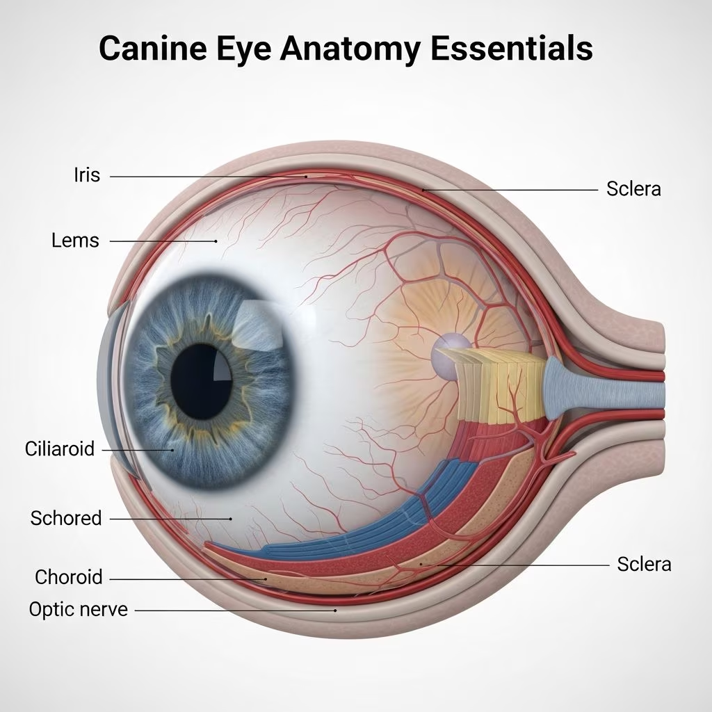
The canine eye is a remarkable organ engineered for survival in diverse environments, featuring adaptations that prioritize motion detection and low-light performance over color acuity or fine detail. Unlike human eyes, dog eyes excel in dim conditions thanks to specialized structures like a larger lens and reflective layers.
Protective Outer Layers of the Dog Eye
Dogs possess a robust outer shell that safeguards the delicate internal components. The outermost visible part is the sclera, a durable white fibrous layer that encases most of the eyeball, providing structural integrity against impacts. Overlying the sclera at the front is the transparent cornea, a curved dome that not only shields the eye but also bends incoming light rays to initiate focus.
A thin, moist membrane known as the conjunctiva lines the inner surfaces of the eyelids and extends over the sclera up to the cornea’s edge. This tissue keeps the eye lubricated and can become inflamed during infections or allergies, appearing pink and irritated. These layers work in tandem to form a barrier against dust, debris, and pathogens.
Dynamic Light Regulation: Iris and Pupil Mechanics
Central to light control is the iris, the pigmented ring surrounding the pupil, which contracts or expands via muscle fibers to adjust light entry. In bright settings, the iris narrows the pupil—the central black aperture—to reduce glare; in darkness, it dilates widely to capture maximal light. Dogs’ pupils can open significantly larger than humans’, enhancing their twilight vision.
This system responds reflexively, ensuring the sensitive inner eye remains protected while optimizing image clarity for the brain.
Focusing Powerhouse: The Lens and Ciliary Body
Positioned behind the iris, the lens is a flexible, crystalline structure that fine-tunes focus by altering shape. Connected to the ciliary body, tiny muscles thicken the lens for near objects and flatten it for distant ones, though dogs have less accommodation range than humans. The ciliary body also secretes aqueous humor, a nutrient-rich fluid filling the eye’s front chamber, maintaining pressure and shape.
This fluid circulates through the pupil and drains via the trabecular meshwork at the cornea-sclera junction, preventing buildup that could lead to glaucoma.
Vision Processing Center: Retina and Photoreceptors
At the eye’s rear lies the retina, a neural tapestry packed with photoreceptors: rods** for dim-light sensitivity and motion, and **cones** for color and brightness. Dogs boast a rod-heavy retina, fueling superior night sight, with a reflective **tapetum lucidum** behind it bouncing light back through cells for a second pass—causing the glow in headlights.
| Feature | Dog Eye | Human Eye |
|---|---|---|
| Lens Size | 3-4x larger | Smaller |
| Pupil Dilation | Larger max opening | Limited dilation |
| Tapetum | Present (reflective) | Absent |
| Rod:Cone Ratio | High rods | Balanced |
This table highlights key differences enabling dogs’ 20/70-20/80 acuity but excels in low light. The **area centralis**, a cone-rich zone, sharpens central vision. Signals converge into the **optic nerve** for brain relay.
Shielding Mechanisms: Eyelids and Third Eyelid
Dogs feature upper and lower eyelids that blink frequently to distribute tears and remove irritants. Uniquely, a third eyelid (nictitating membrane) sits in the medial canthus, sliding horizontally for extra protection during rough play or illness. Normally retracted, it protrudes with inflammation or anesthesia.
- Upper/lower eyelids: Spread tears, block light.
- Third eyelid: Shields cornea, sweeps debris.
Tear Film Dynamics and Gland Functions
A stable tear film—watery base, oily layer, mucin coat—nourishes and defends the cornea. Lacrimal glands above each eye produce aqueous tears; Meibomian glands in lids add lipids; conjunctival goblet cells** secrete mucus. Tears drain via **nasolacrimal ducts** to the nose, explaining watery eyes during emotion.
Imbalances cause dry eye (keratoconjunctivitis sicca), underscoring tear importance.
The Orbit: Eye’s Secure Housing
Housed in the bony **orbit**, the eye gains stability from surrounding skull bones, plus muscles for movement, nerves, vessels, and glands. Extra retractor muscles let dogs pull eyes deep into sockets for safety. This setup allows wide peripheral vision, vital for predators/prey.
Vision Adaptations: Why Dogs See Differently
Dogs’ larger corneas and pupils funnel more light; the tapetum amplifies it. High rod count prioritizes dusk/dawn activity, detecting motion afar but blurring fine print or distant colors (limited to blue/yellow spectra). Visual field spans ~240°, with binocular overlap for depth.
Common Eye Health Indicators
Owners should monitor for redness, squinting, discharge, or cloudiness. Breed predispositions (e.g., brachycephalics’ prominent eyes) heighten risks. Routine vet exams catch issues early.
Frequently Asked Questions (FAQs)
Why do dog eyes glow green at night?
The tapetum lucidum reflects light, enhancing night vision and creating the glow.
Do dogs see color?
Yes, but dichromatic: blues, yellows; reds/oranges appear as grays.
What is the third eyelid for?
Extra protection, lubrication, and debris clearance.
Can dogs see as sharply as humans?
No, ~20/75 vision, but excel in low light and motion.
How often should I check my dog’s eyes?
Daily for abnormalities; vet annually.
Maintaining Optimal Canine Vision
Protect eyes with goggles during high-risk activities, trim facial hair, and ensure UV-safe environments. Nutrition with antioxidants supports retinal health. Prompt vet care prevents minor issues escalating.
In summary, understanding these elements empowers owners to safeguard their pet’s sight, appreciating evolutionary tweaks for pack hunting and survival.
References
- Eye Structure and Function in Dogs — MSD Veterinary Manual. 2023. https://www.msdvetmanual.com/dog-owners/eye-disorders-of-dogs/eye-structure-and-function-in-dogs
- Structure of the Canine Eye — Whole Dog Journal. 2022-10-15. https://www.whole-dog-journal.com/health/structure-of-the-canine-eye/
- Eye Problems in Dogs — PDSA. 2024. https://www.pdsa.org.uk/pet-help-and-advice/pet-health-hub/symptoms/eye-problems-in-dogs
- How Your Dog’s Eyes Differ From Yours — Rex Specs. 2023-05-20. https://www.rexspecs.com/blogs/news/how-your-dogs-eyes-differ-from-yours
- Pet Eye Disease, Dog Eye Anatomy And Structure — Safari Veterinary Care Centers. 2023. https://www.safarivet.com/care-topics/dogs-and-cats/eye-disease/
- Dog Vision and Eye Anatomy: How Dogs See — PetMD. 2024-01-10. https://www.petmd.com/dog/general-health/how-do-dogs-see-world



