

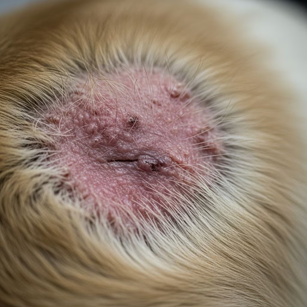
Canine atopic dermatitis represents one of the most prevalent chronic skin disorders in dogs, marked by relentless itching and inflammation triggered by environmental allergens. This condition, often genetically influenced, demands a multifaceted approach to diagnosis and long-term control to enhance the pet’s quality of life.
Understanding the Roots of Atopic Dermatitis in Dogs
At its core, canine atopic dermatitis stems from an exaggerated immune response to common environmental substances. Dogs with this condition produce IgE antibodies against allergens like pollen, dust mites, and molds, leading to skin barrier dysfunction and persistent inflammation. Genetic predisposition plays a pivotal role, with certain breeds such as Golden Retrievers, Labrador Retrievers, French Bulldogs, and West Highland White Terriers showing higher susceptibility.
The skin’s outer layer, or epidermis, in affected dogs is inherently defective, allowing easier penetration of allergens. This initiates a cascade of inflammatory signals, resulting in pruritus—the hallmark symptom. Unlike acute allergies, atopic dermatitis is lifelong, often manifesting in puppyhood or young adulthood, and can wax and wane with seasonal changes or persist year-round.
Recognizing the Signs: Symptoms and Progression
The primary indicator of canine atopic dermatitis is intense pruritus, prompting dogs to chew, lick, and scratch excessively. Common affected areas include the paws, face, ears, armpits, groin, and abdomen. Owners may first notice subtle redness or mild scaling, which progresses if unchecked.
- Early signs: Erythema (redness), small papules, and mild hair loss from rubbing.
- Progressive changes: Excoriations (scratch marks), crusting, lichenification (thickened skin), and hyperpigmentation.
- Complications: Recurrent bacterial infections (pyoderma) from Staphylococcus or yeast overgrowth (Malassezia), leading to odor, oozing, and ear infections.
Symptoms can be seasonal, tied to pollen peaks, or perennial, exacerbated by indoor allergens. Chronic cases may present with salivary staining on paws or fur from constant licking.
Key Differential Diagnoses: Ruling Out Mimics
Accurate diagnosis requires excluding conditions that mimic atopic dermatitis. A systematic workup is essential, starting with history and physical exam.
| Condition | Key Features | Diagnostic Tests |
|---|---|---|
| Flea Allergy Dermatitis | Pruritus at tail base, lumbosacral area, thighs | Flea combing, trial flea control |
| Food Allergy | Year-round itching, GI signs, ears/paws affected | Elimination diet trial (8-12 weeks) |
| Scabies/Mange | Papules, crusting, hair loss on ears/elbows | Skin scrapings, response to acaricides |
| Pyoderma | Pustules, epidermal collarettes, epidermal collarettes | Cytology, skin scrapings, culture |
| Malassezia Dermatitis | Greasy skin, odor, ventral abdomen | Cytology (yeast count >5/HPF) |
Flea infestations must be addressed first, as even atopic dogs can have concurrent flea allergies. Skin cytology and scrapings help identify secondary infections, which often amplify pruritus.
Diagnostic Approach: Steps to Confirmation
Diagnosis hinges on Favrot’s criteria, a validated set of clinical features: pruritus onset before age 3, affecting paws/face/ears, chronicity, and response patterns to therapies. No single test confirms atopy; instead, it’s by exclusion.
- History and Exam: Note lesion distribution, seasonality, breed.
- Rule Out Parasites: Flea comb, trial preventives.
- Manage Infections: Antibiotics/antifungals based on cytology.
- Diet Trial: Hydrolyzed or novel protein diet for perennial cases.
- Allergy Testing: Intradermal (IDT) or serum IgE for immunotherapy candidates.
IDT involves injecting allergens intradermally; positive reactions form wheals. Serum tests detect allergen-specific IgE. Neither is purely diagnostic but guides treatment.
Treatment Strategies: Multi-Modal Management
Control requires addressing itch, infections, and allergens lifelong. No cure exists, but combinations yield excellent results.
Anti-Pruritic and Anti-Inflammatory Options
- Oclacitinib (Apoquel): Rapid itch relief via JAK inhibition; effective within hours, suitable for long-term use.
- Lokivetmab (Cytopoint): Monoclonal antibody targeting IL-31; monthly injections, minimal side effects.
- Cyclosporine (Atopica): Immunosuppressant; takes 4-6 weeks, good for maintenance.
- Glucocorticoids: Potent but short-term due to side effects like polyuria/polydipsia.
Allergen-Specific Immunotherapy (ASIT)
ASIT is the cornerstone for modifying disease, desensitizing via sublingual drops or injections. Testing identifies triggers; response seen in 60-80% of cases after 9-12 months, reducing medication needs.
Topical and Supportive Care
Regular medicated shampoos (chlorhexidine, antifungal) prevent infections. Omega-3 fatty acids and ceramide-enriched diets bolster skin barrier. Environmental control—HEPA filters, frequent bathing—limits exposure.
Breeder Perspectives: Prevention and Early Intervention
For breeders, screening pedigrees for atopy history is crucial. Puppies from affected lines benefit from fortified puppy diets reducing incidence. Early signs warrant prompt vet referral; breeders can educate owners on multimodal plans.
Prognosis and Long-Term Outlook
With diligent management, most dogs achieve good control, minimizing flares. Regular vet monitoring adjusts therapies. Owners report high satisfaction with modern options like biologics and ASIT.
Frequently Asked Questions (FAQs)
What breeds are most prone to atopic dermatitis?
Commonly affected: Shar-Pei, Bulldog breeds, Retrievers, Terriers.
Can diet alone cure atopy?
No, but elimination trials rule out food allergies; supportive nutrition aids skin health.
How effective is immunotherapy?
60-80% success rate in reducing signs long-term.
Is Cytopoint safe for all dogs?
Generally yes, even young puppies; rare hypersensitivity.
How often should I bathe my atopic dog?
1-2 times weekly with vet-recommended products.
References
- Canine atopic dermatitis: detailed guidelines for diagnosis and … — International Committee on Allergic Diseases of Animals (ICADA). 2015-07-22. https://pmc.ncbi.nlm.nih.gov/articles/PMC4531508/
- Understanding Atopic Dermatitis in Dogs: A Breeder’s Guide — MyDogDNA. Accessed 2026. https://mydogdna.com/blogs/news/understanding-atopic-dermatitis-in-dogs-a-breeder-s-guide
- Canine Atopic Dermatitis (Environmental Allergy) in Dogs — MedVet. Accessed 2026. https://www.medvet.com/atopic-disease-dogs/
- A Comprehensive Look at Canine Atopic Dermatitis — Clinician’s Brief. Accessed 2026. https://www.cliniciansbrief.com/custom/canine-atopic-dermatitis
- Canine Atopic Dermatitis — Merck Veterinary Manual. Accessed 2026. https://www.merckvetmanual.com/integumentary-system/atopic-dermatitis/canine-atopic-dermatitis



