

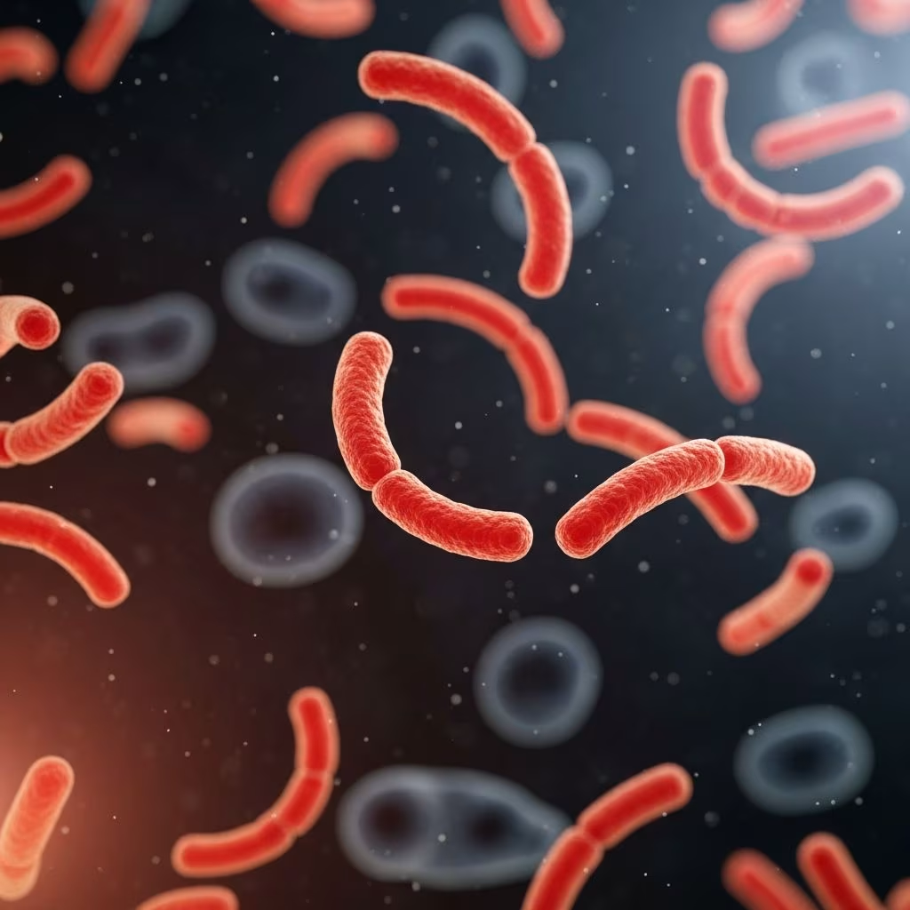
Campylobacteriosis represents a significant bacterial infection affecting the gastrointestinal tracts of various animals, particularly dogs, cats, and livestock. Caused primarily by species like Campylobacter jejuni and Campylobacter upsaliensis, this condition leads to enteritis, manifesting as diarrhea and related symptoms. Understanding its pathology, spread, and control is crucial for veterinarians and pet owners alike.
Understanding the Bacterial Culprits
The genus Campylobacter comprises spiral-shaped, microaerophilic bacteria thriving in low-oxygen environments. C. jejuni stands out as the most common pathogen in canines, often linked to acute enteric disturbances. These bacteria possess flagella for motility, enabling colonization of the intestinal mucosa. In animals, they adhere to epithelial cells, triggering inflammation and disrupting normal gut function.
Other species, such as C. upsaliensis, play roles especially in cats and dogs, while C. coli appears more in livestock. These pathogens are thermophilic, favoring body temperatures around 42°C in animals, which aids their survival in hosts.
Epidemiology and Prevalence Across Species
Campylobacter infections are widespread. Studies indicate up to 49% of dogs may carry the bacteria asymptomatically, shedding them in feces and posing risks to cohorts. In healthy versus diarrheic dogs, prevalence varies significantly, with higher rates in those showing acute symptoms.
A survey in India revealed substantial shedding among dogs attending veterinary clinics, associating it with shared habitats and multi-pet environments. Puppies under six months and stressed adults—due to hospitalization, surgery, or transport—are particularly vulnerable. Livestock like calves and pigs also harbor these bacteria, amplifying farm-to-pet transmission risks.
- Dogs: Highest incidence in young pups; multidrug-resistant strains emerging.
- Cats: Often subclinical but can develop severe diarrhea.
- Livestock: Common in calves, sheep, pigs; contributes to environmental contamination.
Transmission Pathways in Animal Populations
These bacteria spread via the fecal-oral route. Contaminated water, food, or direct contact with infected feces facilitates transmission. Dogs in group settings, such as kennels or parks, face elevated risks from shared bowls or soil.
Raw diets, exposure to wildlife, or contact with livestock heightens susceptibility. Vertical transmission from dam to offspring occurs rarely, but communal living accelerates outbreaks. Zoonotic potential underscores hygiene needs, as identical strains link pet and human cases.
Clinical Manifestations and Symptom Profiles
Symptoms range from mild to severe. Acute cases feature watery, mucoid, or bloody diarrhea lasting 5-15 days, often with anorexia, lethargy, vomiting, and fever. Puppies exhibit bile-streaked stools with leukocytes.
In chronic forms, intermittent loose stools persist. Rare complications include bacteremia, cholecystitis with icterus, and thickened gallbladder walls visible on ultrasound. Cats mirror canine signs but may show less blood in feces.
| Species | Common Symptoms | Severity Factors |
|---|---|---|
| Dogs | Watery/bloody diarrhea, anorexia, fever | Age <6 months, stress |
| Cats | Mucoid stools, lethargy | Concurrent illnesses |
| Livestock | Diarrhea, dehydration | Crowding, poor hygiene |
Pathological Changes in Affected Tissues
Histologically, infections cause mucosal damage: reduced epithelial height, villous stunting, goblet cell depletion, and lamina propria infiltration by inflammatory cells. Hyperplasia thickens the mucosa, with subepithelial hemorrhage and congestion in colon and cecum.
These alterations impair absorption, leading to osmotic diarrhea. In severe cases, Peyer’s patches hyperplasticate, signaling immune response.
Diagnostic Approaches and Laboratory Confirmation
Diagnosis hinges on fecal analysis. Culture on selective media like modified charcoal cefoperazone deoxycholate agar (mCCDA) with supplements excels for isolation. PCR detects DNA rapidly, while microscopy reveals leukocytes or spiral bacteria.
Culture confirms species and sensitivity, vital for persistent cases. Blood/urine tests rule out systemic involvement. Direct plating on Campy-BAP suffices for acute samples; enrichment aids low-shedding carriers.
- Collect fresh feces.
- Selective culture/PCR.
- Sensitivity testing for resistance profiling.
Treatment Strategies and Antimicrobial Options
Mild cases often resolve spontaneously, but severe ones demand intervention. Erythromycin or tylosin target C. jejuni and C. upsaliensis, though resistance is rising. Supportive care includes fluids, antiemetics, and isolation.
Gentamicin and amikacin show in vitro efficacy, but enrofloxacin risks contraindications in dogs. Post-treatment shedding may persist, indicating carrier states.
Prevention Measures for Herds and Households
Hygiene is paramount: clean water, cooked diets, and waste isolation curb spread. Avoid raw meat; quarantine new animals. Probiotics may bolster gut flora, though evidence is preliminary.
Farm biosecurity—disinfection, rodent control—limits livestock reservoirs. Pet owners should wash hands post-handling, especially with children or immunocompromised individuals.
Zoonotic Implications and Public Health Concerns
C. jejuni causes human gastroenteritis; pets contribute via shared strains. Multidrug-resistant isolates from dogs link to outbreaks, urging surveillance. Pet ownership elevates human risk, particularly with diarrhea.
Research Frontiers and Future Directions
Ongoing studies probe prevalence, resistance via whole-genome sequencing, and risk factors like diet or antibiotics. Enhanced diagnostics promise better control.
Frequently Asked Questions (FAQs)
What causes Campylobacteriosis in dogs?
Bacteria like C. jejuni, transmitted fecal-orally.
Is Campylobacter contagious to humans?
Yes, zoonotic; practice hygiene.
How long does diarrhea last?
5-15 days untreated; antibiotics shorten.
Can healthy pets carry it?
Yes, up to 49% asymptomatically.
What antibiotics work best?
Erythromycin, tylosin; test sensitivity.
References
- Multidrug-Resistant Campylobacter Jejuni in Dogs; Underdiagnosed Cause of Enteric Disease — Cornell University College of Veterinary Medicine. 2021-08. https://www.vet.cornell.edu/research/awards/202108/multidrug-resistant-campylobacter-jejuni-dogs-underdiagnosed-cause-enteric-disease
- Bacterial Infection (Campylobacteriosis) in Dogs — PetMD. Accessed 2026. https://www.petmd.com/dog/conditions/digestive/c_multi_campylobacteriosis
- Enteric Bacterial Infections — Veterian Key. Accessed 2026. https://veteriankey.com/enteric-bacterial-infections/
- Campylobacter Infection in Dogs — VCA Animal Hospitals. Accessed 2026. https://vcahospitals.com/know-your-pet/campylobacter-infection-in-dogs
- Prevalence, associated risk factors and antimicrobial susceptibility of Campylobacter species from dogs — PMC (PubMed Central). 2020-07-22. https://pmc.ncbi.nlm.nih.gov/articles/PMC7386756/
- Zoonotic Prevention Flyer: Campylobacteriosis — Pet Advocacy Network. 2022-02. https://petadvocacy.org/wp-content/uploads/2022/02/Zoonotic_Prevention-Flyer-Campylobacter.pdf
- Campylobacter culture in cats and dogs – money well spent? — Awanui Vets. 2019-09. https://www.awanuivets.co.nz/wp-content/uploads/2019/09/Campylobacter-culture-in-cats-and-dogs-money-well-spent-May18.pdf



