

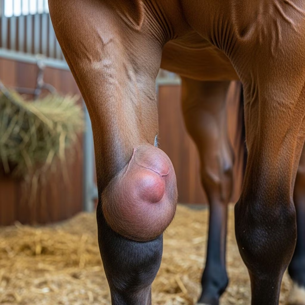
Bursitis represents a significant musculoskeletal challenge in large animals such as horses, cattle, and pigs, characterized by inflammation of the bursa—a fluid-filled sac that cushions joints and tendons. This condition arises from various triggers including repetitive trauma, direct injury, or bacterial invasion, leading to swelling, pain, and potential lameness that can impair productivity and welfare. Understanding its pathology, clinical presentations, and management is crucial for veterinarians and livestock managers aiming to mitigate economic losses and improve animal outcomes.
Understanding the Anatomy and Function of Bursae
Bursae are synovial-lined sacs strategically located to minimize friction between tendons, muscles, bones, and skin during movement. In large animals, they are particularly vital in high-motion areas like elbows, hocks, and shoulders. Natural or ‘true’ bursae exist congenitally deep within tissues, while ‘acquired’ ones form subcutaneously due to chronic pressure. Inflammation disrupts this lubricating role, causing fluid accumulation, wall thickening, and fibrosis, which can progress to chronic, non-painful enlargements or acute, debilitating infections.
In horses, common sites include the olecranon (capped elbow or shoe boil), tuber calcaneus (capped hock), and supraspinous region (fistulous withers). Cattle often develop tarsal bursitis from lying on hard surfaces, while pigs show adventitious bursae on hocks from similar environmental factors. These adaptations highlight how bursae respond to mechanical stress, but persistent irritation leads to pathological changes.
Primary Causes and Risk Factors
Bursitis etiology spans mechanical, infectious, and idiopathic origins. Repetitive trauma is paramount: in horses, shoe contact causes olecranon bursitis; stall kicking leads to capped hocks. Cattle tarsal lesions stem from abrasive cubicle bedding or concrete floors, with prevalence up to 71% in poor housing. Pigs exhibit 51% hock bursitis prevalence, varying by unit from 10-84%, linked to flooring.
- Trauma-induced: Chronic pressure forms acquired bursae; acute injuries cause hematomas that organize into inflammatory sacs.
- Infectious: Bacterial entry via wounds or hematogenous spread, with Brucella spp. implicated in fistulous withers.
- Species-specific risks: Larger cattle have higher tarsal incidences due to weight; thin body condition exacerbates lying time and pressure.
Environmental factors amplify risks: dirty, slippery floors increase falls; inadequate bedding promotes abrasions. Behavioral issues, like stall vices in horses, compound mechanical stress.
Clinical Manifestations Across Species
Symptoms vary by stage and location. Acute bursitis presents with hot, painful, fluctuant swellings, heat, and lameness. Chronic forms evolve into firm, painless masses with thickened walls, excess fluid, and fibrous septa, rarely causing gait issues unless massive.
| Species | Common Site | Key Signs | Prevalence/Notes |
|---|---|---|---|
| Horses | Olecranon (shoe boil) | Fluid-filled elbow swelling from shoe trauma | Common in stabled horses |
| Horses | Tuber calcaneus (capped hock) | Dorsal hock enlargement from kicking | Behavioral modification key |
| Cattle | Tarsal/hock | Lateral swelling, possible hair loss, cellulitis | 50-71% in cubicles |
| Pigs | Hock aspects | Mild lateroplantar bursae most common | 51% overall |
Septic cases escalate with abscessation, fistulae, and severe lameness. In fistulous withers, straw-colored exudate progresses to suppuration post-rupture, involving vertebral structures.
Diagnostic Approaches
Diagnosis relies on history, physical exam, and imaging. Palpation reveals fluctuant or firm swellings; lameness assessment differentiates from joint issues. Ultrasonography confirms fluid, wall thickness, and septations, distinguishing bursitis from abscesses or tumors. Aspiration yields synovial fluid for cytology, culture, and Brucella titers in suspect cases.
- History: Trauma exposure, housing, behavior.
- Exam: Localized edema, heat, pain on pressure.
- Advanced: Radiography for bone involvement; bursoscopy for endoscopy.
Differential diagnoses include synovial effusions, cellulitis, or neoplasia; exclusion via imaging is standard.
Management and Treatment Strategies
Treatment tiers from conservative to surgical, guided by acuity and infection.
Conservative Therapies
Early acute cases benefit from cold hydrotherapy to reduce inflammation, followed by aseptic aspiration and corticosteroid injection. Counterirritants, ultrasound, or radiation aid chronic non-septic hygromas. Shoe-boil rolls prevent recurrence in elbows; hock boots or stall redesign curb trauma.
Surgical Interventions
Refractory or infected bursae demand surgery: curettage, drainage, or complete excision (bursectomy). Arthroscopic bursoscopy minimizes invasion in horses. Septic cases require systemic antibiotics post-drainage, with daily lavage. In cattle purulent tarsal bursitis, fistulae enlargement facilitates pus evacuation. Presternal bursae in large animals respond to lancing and dressing.
Prognosis excels in early, non-septic cases; chronic infections like fistulous withers often necessitate euthanasia due to cost and recurrence.
Prevention in Farm and Stable Settings
Proactive measures slash incidence. Optimal housing is foundational: deep, absorbent bedding in cattle cubicles reduces tarsal trauma; non-slip, soft flooring for pigs. Horse stables should feature kick-proof walls, adequate space, and protective gear like hock boots.
- Housing redesign: Rubber mats, rounded edges.
- Bedding management: Frequent renewal, dirt as potential protectant despite infection risk.
- Behavioral: Enrichment to deter vices; body condition monitoring.
- Farriery: Proper shoeing avoids elbow contact.
Herd-level changes yield swift improvements; larger cows benefit from weight management.
Species-Specific Insights
Horses: Elbow and Hock Focus
Olecranon bursitis (shoe boils) forms from heel bulb pressure; surgical excision under sedation removes intact sacs. Capped hocks resolve with behavioral fixes; excision for persistent cases.
Cattle: Tarsal Challenges
Chronic cellulitis from pressure; uncomplicated cases self-limit with housing tweaks. Infection risks arthritis.
Pigs: Hock Bursitis Prevalence
Mild lesions predominate; unit variations underscore flooring importance.
Frequently Asked Questions (FAQs)
What is the most common cause of bursitis in horses?
Repetitive trauma, such as shoe contact with elbows or kicking stalls.
Can bursitis in cattle lead to lameness?
Yes, severe cases with infection cause significant gait issues and joint involvement.
Is surgery always required for shoe boils?
No, small, non-painful ones may be managed conservatively; surgery for large or infected.
How can farmers prevent tarsal bursitis?
Improve bedding quality, use soft flooring, and monitor cow comfort.
What is the prognosis for septic bursitis?
Guarded; early intervention improves outcomes, but chronic cases may recur.
References
- Bursitis in Large Animals – Musculoskeletal System – Veterinary Manual — Merck Veterinary Manual. 2020. https://www.merckvetmanual.com/musculoskeletal-system/bursitis-in-large-animals/bursitis-in-large-animals
- Adventitious bursitis of the hock in finishing pigs — PubMed (Veterinary Record). 1998-04-11. https://pubmed.ncbi.nlm.nih.gov/9501386/
- Tarsal bursitis and cellulitis in Cows — Vetlexicon Bovis. Accessed 2026. https://www.vetlexicon.com/bovis/dermatology/articles/tarsal-bursitis-and-cellulitis/
- Shoe Boils (Olecranon Bursitis or Capped Elbow) in Horses — Mad Barn. Accessed 2026. https://madbarn.com/shoe-boils-in-horses/
- Alexandria Journal of Veterinary Sciences on Presternal Bursitis — Alexandria J Vet Sci. 2017. https://www.alexjvs.com/index.php?fulltxt=267069&fulltxtj=31&fulltxtp=31-1495049902.pdf
- Morphoclinical Aspects in Tarsal Bursitis in Cattles — CABI Digital Library. 2008. https://www.cabidigitallibrary.org/doi/pdf/10.5555/20083298016



