

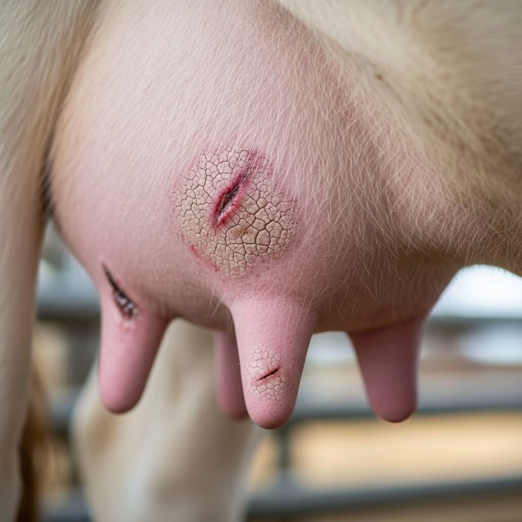
The udder represents a cornerstone of dairy production, serving as the primary site for milk synthesis and storage in cows. Composed of four independent quarters, each with its own teat, the bovine udder relies on a complex network of ligaments, tissues, and vascular structures for support and function. Disruptions from physical trauma or inherent structural flaws can severely impair lactation, lead to secondary infections, and compromise overall animal welfare. This article delves into the anatomy basics, common injury types, diagnostic approaches, therapeutic interventions, and proactive prevention measures to equip farmers and veterinarians with actionable knowledge.
Fundamentals of Bovine Udder Anatomy
Understanding udder anatomy is essential for identifying disorders. The mammary gland consists of parenchyma, the milk-producing tissue made of alveoli and ducts, and stroma, which includes connective, vascular, and adipose elements. Each quarter functions autonomously, preventing milk crossover between sides. The udder suspends from the abdominal wall via a multi-layered system: superficial skin and fascia provide minimal hold, while deeper coarse areolar tissues anchor front quarters.
Key suspensory components include the subpelvic tendon, originating from pelvic attachments and giving rise to lateral ligaments. These fibrous bands, with limited elasticity, wrap around udder halves without uniting centrally, allowing balanced suspension under heavy milk loads up to 50 kg. The median suspensory ligament, formed by elastic yellow sheets, divides halves and stretches to accommodate filling, ensuring stability at the udder’s center of gravity.
- Skin and superficial fascia: Basic coverage with minor support.
- Coarse areolar tissue: Bonds front quarters to body wall.
- Lateral suspensory ligaments (superficial and deep): Fibrous extensions enveloping sides.
- Median suspensory ligament: Elastic core for balanced hold.
Milk pathways feature gland cisterns (100-400 ml storage), teat cisterns, and sphincters protected by keratin plugs, barring microbial entry until parturition.
Common Traumatic Injuries to the Udder
Trauma arises from environmental hazards, handling errors, or machinery mishaps in dairy operations. Front teats, more exposed, and rear ones in buffaloes (analogous risks in cows) face higher injury rates due to positioning.
Teat and Udder Lacerations
Sharp objects like gates, fences, or broken glass cause lacerations. Superficial cuts heal readily with cleaning and bandaging, but deep wounds penetrating cisterns risk mastitis from bacterial invasion. Symptoms include swelling, hemorrhage, and milk leakage. Immediate irrigation with antiseptics and suturing under sedation prevent complications.
Blunt Force Contusions
Kicks from herd mates or falls during slips produce bruising. Hematomas form under skin, causing pain and reduced milk yield. Cold therapy initially reduces swelling, followed by warm compresses for resolution. Monitor for abscessation.
Milking Equipment Trauma
Vacuum fluctuations or ill-fitted liners induce ‘teat end cracks’ or hyperkeratosis. Chronic irritation erodes sphincter integrity, inviting pathogens. Regular equipment calibration mitigates this.
| Injury Type | Common Causes | Immediate Signs | Risk Factors |
|---|---|---|---|
| Lacerations | Sharp farm hazards | Bleeding, wounds | Overcrowded pens |
| Contusions | Trauma impacts | Swelling, lameness | Slippery floors |
| Equipment damage | Milking machine faults | Cracks, lesions | Poor maintenance |
Structural Weaknesses and Suspensory Failures
Developmental or age-related defects undermine udder integrity. Weak ligaments lead to pendulous udders, increasing trauma susceptibility and mastitis odds.
Median Ligament Weakness
This elastic divider, crucial for balance, degrades from repeated stretching or genetic predisposition. Affected udders sag centrally, with quarters separating excessively. Early signs: visible groove deepening during lactation. Severe cases (‘cleft udder’) hinder milking and predispose to injuries.
Lateral Ligament Insufficiency
Fibrous lateral bands fail to envelop properly, causing sideward splaying. High-producing breeds like Holsteins, bearing 50-100 kg udders, strain these non-elastic structures. Bilateral weakness results in ‘airplane udder’ conformation, elevating mastitis risk via poor drainage.
Quarter Asymmetry and Edema
Imbalanced growth or post-trauma fibrosis yields uneven quarters. Chronic edema from vascular compromise swells tissues, impairing function. Diuretics and massage aid mild cases; surgery addresses fibrosis.
- Genetic screening: Select sires with strong udder composites.
- Age monitoring: Older cows (>5 lactations) prone to ligament tears.
- Nutritional support: Adequate selenium/vitamin E prevents edema.
Diagnostic Techniques for Udder Disorders
Accurate diagnosis combines visual exams, palpation, and imaging. Observe during milking for leakage or asymmetry. Palpate ligaments for tears—median feels like taut elastic, laterals as firm cables.
Ultrasound detects cistern integrity, hematomas, or abscesses. Milk cultures identify infections secondary to trauma. California Milk Cell Test quantifies inflammation.
- Visual inspection: Check for scars, swellings.
- Palpation: Assess ligament tension.
- Imaging: Ultrasound for internal damage.
- Lab tests: Cultures, cytology.
Treatment Strategies for Udder Trauma and Defects
Intervene promptly to preserve quarters. Clean wounds with saline, apply topical antimicrobials. Systemic antibiotics guard against mastitis.
For suspensory failures, supportive slings temporarily elevate pendulous udders. Surgical ligament repair, though rare, uses grafts in elite animals. Pain management with NSAIDs improves mobility.
Regenerative therapies like stem cell injections show promise for ligament healing, backed by veterinary trials.
Prevention Best Practices in Dairy Herds
Proactive management minimizes risks. Design barns with rounded edges, non-slip flooring. Train staff on gentle handling.
Breeding programs prioritize udder conformation scores: strong attachments score 8+ on linear scales. Routine hoof trimming prevents kicks.
Milking protocols: Pre-dip teats, ensure vacuum stability (42-45 kPa). Nutritional balance supports tissue resilience—adequate energy prevents excessive udder fill straining ligaments.
| Prevention Measure | Benefit | Implementation Tip |
|---|---|---|
| Barn modifications | Reduces trauma | Pad gates, rubber mats |
| Genetic selection | Stronger structures | Use EPDs for udder traits |
| Milking hygiene | Prevents cracks | Daily liner checks |
| Nutrition | Tissue health | Balanced rations |
Impact on Production and Economics
Udder disorders slash yields: a traumatized quarter drops 20-50% output. Mastitis from breaches costs $200-400/case in treatments and lost milk. Culling weak-uddered cows averages $1,500/head.
Herd-level, 10% prevalence halves profitability. Early intervention recovers 80% function versus permanent loss.
FAQs on Bovine Udder Health
What causes a pendulous udder in cows?
Primarily ligament weakening from genetics, age, or high yield stress.
How do I treat a torn teat?
Clean, suture if deep, antibiotics, and rest from milking.
Can structural defects be bred out?
Yes, via genomic selection for suspensory strength.
Is surgery common for udder injuries?
Rare; conservative care suffices for most cases.
How often should udders be inspected?
Daily pre-milking, with vet checks quarterly.
References
- Sheep and Goat Udder Basics: Anatomy & Physiology — About Small Ruminants. 2023. https://aboutsmallruminants.com/sheep-goat-udder-basics-anatomy-physiology/
- Anatomy and Affection of Udder and Teat — University of Basrah Faculty. 2023-05-10. https://faculty.uobasrah.edu.iq/uploads/teaching/1683370598.pdf
- Anatomy of Mammary Gland — Groupe ESA. 2023. https://www.groupe-esa.com/ladmec/bricks_modules/brick01/co/ZBO_Brick01_2.html
- Anatomy of the Mammary Gland — Cornell Cooperative Extension. 2023. https://nydairyadmin.cce.cornell.edu/uploads/doc_113.pdf
- The Anatomy of the Dairy Cow Udder — YouTube (Wisconsin Veterinary Diagnostic Lab). 2023. https://www.youtube.com/watch?v=5p4_OLayzlA
- Udder Anatomy and Physiology — University of Minnesota Large Animal Surgery. 2023. https://open.lib.umn.edu/largeanimalsurgery/chapter/udder-anatomy/



