

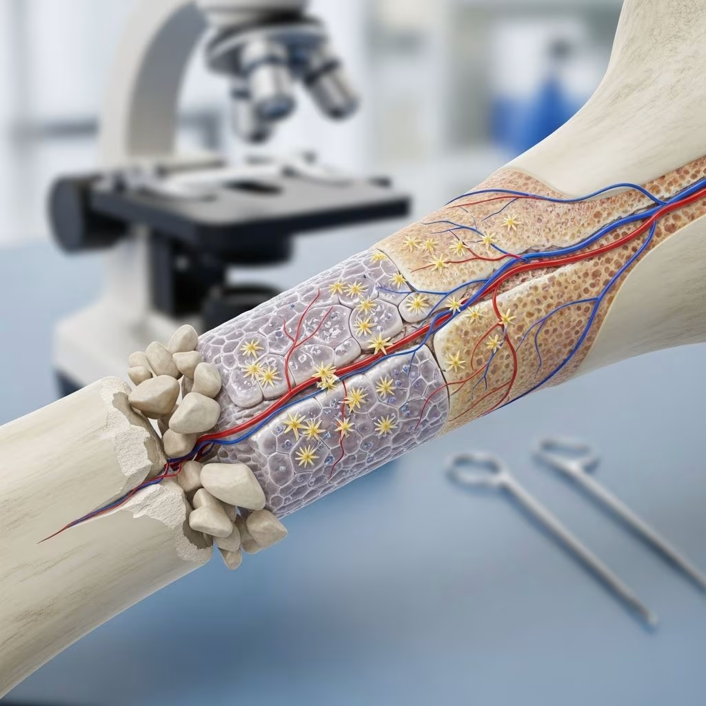
Dogs possess remarkable natural abilities to regenerate bone after fractures or injuries, following a structured healing process that mirrors human physiology but adapted to their active lifestyles. This article explores the mechanisms of bone regeneration in dogs, common injuries, treatment options, and emerging regenerative therapies supported by veterinary research.
How Do Dogs Heal Broken Bones?
Bone healing in dogs occurs through a well-orchestrated biological process involving inflammation, repair, and remodeling phases. Immediately after a fracture, a hematoma forms at the site, triggering an inflammatory response that recruits cells essential for repair. Over weeks, soft callus transitions to hard callus as osteoblasts produce new bone matrix, which is then remodeled into strong, lamellar bone capable of withstanding mechanical stress.
This process typically takes 6-12 weeks for simple fractures in healthy dogs, influenced by factors like age, nutrition, and fracture stability. Studies show that vascular supply is critical, as revascularized bone grafts heal faster than avascular ones, with histological evidence of superior cortical repair and regular bone plate arrangement by 12 weeks post-implantation.
- Inflammation phase (days 1-5): Blood clot formation and immune cell infiltration clear debris.
- Repair phase (weeks 1-6): Fibrocartilaginous callus bridges the fracture gap.
- Remodeling phase (months 3+): Bone reshapes to original contour via osteoclast and osteoblast activity.
Types of Bone Fractures in Dogs
Dogs are prone to fractures due to high-energy trauma like car accidents, falls, or rough play, especially in breeds with long legs or high activity levels such as Greyhounds or Labrador Retrievers. Fractures are classified by pattern and location:
| Type | Description | Common Sites | Healing Challenge |
|---|---|---|---|
| Simple | Clean break with two fragments | Radius, femur | Low; stable fixation |
| Comminuted | Multiple fragments | Tibia, humerus | High; requires grafting |
| Open | Exposed to environment | Any long bone | Infection risk |
| Non-union | Failure to heal after 6 months | Radius/ulna | Needs biologics |
Non-union fractures, affecting up to 10% of cases, pose significant challenges as they resist conventional fixation, often requiring advanced interventions.
Diagnosis of Bone Injuries
Veterinarians diagnose fractures via physical exams, palpation for crepitus, and imaging. Radiographs reveal fracture lines, displacement, and callus formation, while CT scans provide 3D detail for complex cases. Blood tests monitor bone markers like osteocalcin (OC), bone-specific alkaline phosphatase (BSAP), and procollagen peptides (PICP, PINP), which peak during active healing and confirm progress.
Early diagnosis prevents complications like malunion or infection, with serial X-rays tracking callus maturation from soft to bony density.
Treatment Options for Fractures
Treatment prioritizes anatomical reduction, stable fixation, and biological enhancement. Conservative management suits stable, non-displaced fractures in small dogs, using splints or casts for 4-6 weeks. Surgical options dominate for most cases:
- Internal fixation: Plates, screws, and intramedullary pins provide rigid stability, promoting primary bone healing without callus.
- External fixators: Adjustable frames ideal for open fractures or infections.
- Bone grafting: Autogenous cancellous grafts from iliac crest fill defects and accelerate union.
Post-op care includes crate rest, pain management, and physiotherapy to restore function. Healing is monitored via radiographs at 2, 6, and 12 weeks.
Regenerative Therapies: Accelerating Bone Healing
Advanced therapies harness biologics to enhance natural regeneration, particularly for delayed unions or defects. These include growth factors, stem cells, and scaffolds, showing superior outcomes in peer-reviewed studies.
Bone Morphogenetic Proteins (BMPs)
BMPs, especially recombinant human BMP-2 (rhBMP-2), are potent osteoinductive proteins that recruit mesenchymal stem cells, promote osteoblast differentiation, and stimulate angiogenesis. Applied via carriers or scaffolds at fracture sites, they significantly enhance healing in chronic non-unions. Veterinary use at UC Davis since 2012 has regrown leg bones in dogs with severe defects, removing dead bone and implanting BMP scaffolds for full regeneration. Limitations include cost and immunogenicity, restricting routine use.
Stem Cell Therapy
Mesenchymal stem cells (MSCs) from bone marrow or adipose tissue differentiate into osteoblasts, secreting growth factors. Injected or scaffold-delivered, MSCs improve callus formation and vascularization, ideal for critical-sized defects.
Platelet-Rich Plasma (PRP) and Hydroxyapatite (HAp)
PRP concentrates growth factors like PDGF and TGF-β, combined with HAp nanoparticles for osteoconduction. In a tibial fracture case, PRP-HAp inoculation led to rapid callus consolidation within 10 days, full functional recovery by 8 weeks, and increased bone density on X-rays. This minimally invasive approach stimulates neoangiogenesis and osteoinduction.
Vascularized Bone Grafts
Revascularized autografts maintain blood supply, outperforming cryopreserved grafts. At 12 weeks, treated dogs showed complete cortical repair, regular bone plates, higher Lane-Sandhu scores, and elevated bone markers, confirming faster healing.
Nutrition and Supplements for Bone Health
Optimal nutrition supports regeneration: high-quality protein, balanced calcium-phosphorus ratios (1.2:1 to 1.8:1), and omega-3 fatty acids reduce inflammation. Supplements like glucosamine aid cartilage repair, while vitamin D and K2 enhance mineralization. Avoid excesses to prevent fibrous osteodystrophy.
- Calcium: 1-1.5% DM for adults, higher for healing.
- Vitamin C: Antioxidant for collagen synthesis.
- Trace minerals: Copper, manganese for enzymes.
Post-Treatment Rehabilitation
Rehab protocols include passive range-of-motion exercises, underwater treadmill, and controlled walks progressing from 5-30 minutes daily. Hydrotherapy builds muscle without joint stress. Monitor for lameness; full return to activity takes 3-6 months.
When to See a Veterinary Orthopedist
Consult specialists for open/commuted fractures, non-unions, or failed initial treatments. Board-certified surgeons offer expertise in regenerative techniques and custom implants.
Prevention of Bone Injuries
Prevent fractures with weight management, joint supplements, non-slip flooring, and breed-specific exercise. Spay/neuter reduces osteosarcoma risk in large breeds; regular wellness checks catch metabolic issues early.
Frequently Asked Questions (FAQs)
What is the average healing time for a dog’s broken bone?
Simple fractures heal in 6-8 weeks; complex cases with biologics may take 8-12 weeks, monitored by X-rays.
Can dogs fully recover from severe bone fractures?
Yes, with proper treatment including BMP or PRP, full function returns, as seen in regrowth cases at UC Davis.
Are there natural ways to speed up bone healing?
Nutrition rich in calcium, vitamins, and anti-inflammatories supports healing; avoid over-supplementation.
What breeds are prone to bone fractures?
Active breeds like Labs, Shepherds, and sight hounds due to speed and jumps.
Is surgery always needed for dog fractures?
No, stable fractures in small dogs can heal conservatively, but surgery is standard for most.
References
- In dogs does the use of bone morphogenetic proteins with internal fixation enhance the rate of healing of fractures compared to internal fixation alone? — Veterinary Evidence. 2024. https://veterinaryevidence.org/index.php/ve/article/download/722/version/575/1149
- Therapeutic Effects of Revascularisation on the Healing of Bone Defects Caused by Open Fracture in Dogs. — PMC/NIH. 2020-03-17. https://pmc.ncbi.nlm.nih.gov/articles/PMC7105986/
- UC Davis Veterinary Orthopedic Surgeons Regrow Dog’s Leg Bone. — UC Davis School of Veterinary Medicine. 2023. https://www.vetmed.ucdavis.edu/news/uc-davis-veterinary-orthopedic-surgeons-regrow-dogs-leg-bone
- Regenerative treatment of canine osteogenic lesions with Platelet Rich Plasma and hydroxyapatite nanoparticles. — Frontiers in Veterinary Science. 2024. https://www.frontiersin.org/journals/veterinary-science/articles/10.3389/fvets.2024.1459714/full
- Orthopedic Surgeons Successfully Utilize Bone Regrowth Technology. — UC Davis School of Veterinary Medicine. 2023. https://www.vetmed.ucdavis.edu/news/orthopedic-surgeons-successfully-utilize-bone-regrowth-technology



