
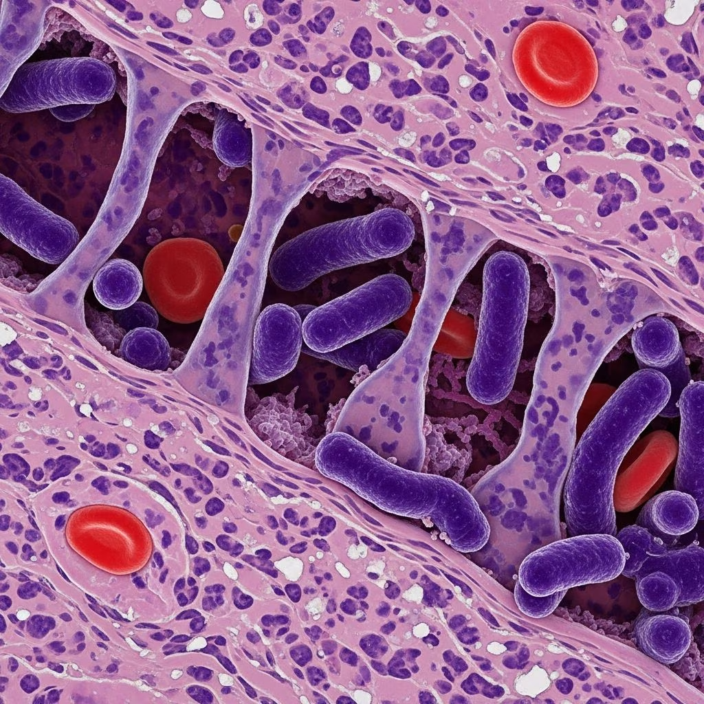
Clostridial bacteria represent a significant threat to animal health across multiple species, from neonatal foals to adult livestock. These gram-positive, anaerobic microorganisms produce powerful toxins that can devastate the gastrointestinal tract, leading to severe morbidity and mortality if not promptly addressed. This comprehensive guide explores the mechanisms of clostridial disease, clinical manifestations, diagnostic strategies, and contemporary management approaches in veterinary medicine.
Pathogenic Mechanisms and Toxin Profiles
Clostridial infections operate through a sophisticated arsenal of toxins that damage intestinal tissue and systemic organs. Understanding these mechanisms is fundamental to recognizing disease patterns and predicting clinical outcomes. Different bacterial strains produce distinct toxin profiles, which directly correlate with disease severity and organ involvement.
Alpha Toxin (CPA) and Intestinal Pathology
Alpha toxin, produced primarily by Clostridium perfringens type A, functions as a phospholipase that disrupts cellular membranes throughout the gastrointestinal tract. This toxin inhibits water absorption in the small intestine, leading to fluid accumulation and characteristic diarrheal symptoms. Experimental models demonstrate that alpha toxin causes multifocal neutrophil infiltration in intestinal tissue layers, creating inflammatory lesions that compromise epithelial integrity.
Beyond intestinal damage, alpha toxin enters systemic circulation in severe infections, producing secondary complications including intravascular hemolysis, platelet aggregation abnormalities, hepatic necrosis, and cardiac dysfunction. This systemic involvement explains the cardiovascular collapse observed in rapidly progressing cases.
Beta Toxin (CPB) and Hemorrhagic Disease
Beta toxin, produced by Clostridium perfringens types B and C, represents one of the most lethal bacterial toxins affecting animals. This toxin specifically targets vascular endothelial cells, causing acute endothelial degeneration and vascular necrosis that compromises blood vessel integrity. The result is catastrophic hemorrhage throughout affected tissues, particularly in the intestinal mucosa and lamina propria.
Type B infections typically manifest as hemorrhagic dysentery in sheep, often progressing to sudden death within 48 hours. Type C infections produce necrotic enteritis characterized by complete loss of absorptive cells along intestinal villi and coagulation necrosis of deeper tissue layers. The rapid progression and high mortality of beta toxin-mediated disease reflects the profound vascular damage this toxin inflicts.
Epsilon Toxin (ETX) and Enterotoxemic Syndromes
Clostridium perfringens type D produces epsilon toxin, which causes enterotoxemia primarily in sheep and goats. This toxin operates through a distinct mechanism involving increased vascular permeability and fluid accumulation in organs, particularly affecting the central nervous system and kidneys. The clinical manifestations of type D enterotoxemia can be replicated through intravenous toxin administration alone, confirming its primary role in disease pathogenesis.
Clinical Manifestations Across Animal Species
Clostridial infections present differently depending on the host species, animal age, bacterial type, and tissue distribution of infection. Recognition of these species-specific patterns enables rapid diagnosis and appropriate therapeutic intervention.
Equine Presentations
Young foals experience particularly severe disease manifestations. Clostridium perfringens causes acute enterocolitis within the first week of life, characterized by inflammation affecting both small and large intestinal segments. Clinical signs include severe abdominal pain and colic, watery diarrhea, progressive dehydration, lethargy, anorexia, and abdominal distention.
The disease progression in foals is notably rapid, with mortality rates exceeding 50% despite intensive medical intervention. Types A and C represent the most common equine pathogens, with type C associated with higher fatality rates due to beta toxin production. Exposure typically occurs through ingestion of contaminated feces in the foaling environment.
Ovine and Caprine Manifestations
Sheep and goats display distinct clinical syndromes depending on bacterial type. Type B infections produce sudden hemorrhagic dysentery accompanied by acute neurological signs, including ataxia and seizure activity. Type C disease manifests as necrotic enteritis with similar acute neurological complications. Type D enterotoxemia presents with neurological symptoms predominating, including depression, seizures, and opisthotonus in peracute cases.
Canine Gastrointestinal Disease
Dogs experience clostridial infections with variable clinical severity. Diarrhea represents the primary clinical sign, with some animals presenting with hemorrhagic fecal material. Affected dogs demonstrate diarrhea rates five times higher in positive cases compared to healthy controls. Interestingly, prior antibiotic therapy is not consistently associated with canine clostridial disease, unlike human infections, though antimicrobial exposure may still represent a risk factor in some populations.
Bacterial Classification and Strain Identification
Clostridial species are classified into five types based on their toxin production profiles. This taxonomic approach provides critical information for predicting disease severity and selecting appropriate interventions.
| Bacterial Type | Primary Toxins | Primary Animal Hosts | Clinical Disease Pattern |
|---|---|---|---|
| Type A | Alpha toxin | Multiple species | Enteritis, gas gangrene, enterotoxemia |
| Type B | Alpha, beta toxins | Sheep, cattle | Hemorrhagic dysentery, acute neurological signs |
| Type C | Alpha, beta toxins | Livestock, humans | Necrotic enteritis, enterotoxemia |
| Type D | Epsilon toxin | Sheep, goats, cattle | Enterotoxemia with neurological manifestations |
| Type E | Iota toxin, beta-2 toxin | Various species | Variable enteritis patterns |
Gas Gangrene and Soft Tissue Infections
Beyond intestinal disease, Clostridium perfringens type A causes gas gangrene (malignant edema) in domestic mammals including sheep, cattle, goats, and horses. This condition typically affects subcutaneous tissue and muscle following traumatic wounds or surgical procedures. The infection progresses rapidly, with tissue necrosis and gas production creating crepitus and severe systemic toxicity.
Gangrenous mastitis represents an additional manifestation of type A infection in cattle and sheep, where the bacteria colonize mammary glands and produce localized necrosis despite systemic toxin production. These soft tissue infections often involve multiple clostridial species acting synergistically to overwhelm tissue defenses.
Diagnostic Approaches and Laboratory Confirmation
Accurate diagnosis of clostridial infections combines clinical presentation with laboratory confirmation. However, bacterial isolation and toxin detection present unique challenges in clinical practice.
Microbiological Culture and Identification
Anaerobic culture of fecal samples or tissue specimens remains the gold standard for bacterial identification. Selective culture media, including blood agar plates with selective supplements, facilitate isolation from mixed microbial populations. Gram staining and biochemical testing characterize the isolated organisms.
Toxin Detection Methods
Polymerase chain reaction (PCR) amplification of toxin genes provides rapid identification of toxigenic strains and specific bacterial types. Diarrheic dogs tested positive for toxigenic Clostridioides difficile in 8.7% of confirmed cases, with toxins A and B detected in fecal samples. Similarly, toxin-encoding genes for Clostridium perfringens can be detected in clinical specimens.
Immunological Assays
Enzyme-linked immunosorbent assays (ELISA) detect specific toxins in fecal or serum samples. These immunological approaches prove particularly valuable in rapid diagnosis, as culture results may require several days to obtain.
Risk Factors and Epidemiological Considerations
Multiple host and environmental factors influence clostridial disease susceptibility and transmission patterns. Understanding these risk factors enables implementation of preventive strategies.
- Neonatal age and immunological immaturity
- Antimicrobial therapy disrupting normal intestinal flora
- Proton pump inhibitor administration altering gastric pH
- Contaminated feeding environments and fecal-oral transmission
- Co-infection with other enteropathogens
- Overcrowding and poor sanitation conditions
- Dietary changes and feed quality
- Prior contact with infected animals
Interestingly, antibiotic exposure presents a paradoxical relationship with clostridial disease. While antimicrobial therapy disrupts protective intestinal microbiota in some cases, many animals with clostridial infections lack recent antibiotic exposure, suggesting alternative pathogenic pathways.
Treatment Protocols and Supportive Care
Managing clostridial infections requires aggressive supportive care combined with specific antimicrobial and immunological interventions. Early recognition and intensive management significantly improve survival outcomes.
Antimicrobial Therapy
Antimicrobials targeting anaerobic bacteria form the cornerstone of treatment. Penicillins, cephalosporins, and metronidazole demonstrate efficacy against clostridial species. Selection depends on local resistance patterns and organ penetration requirements. However, antibiotics alone cannot eliminate toxins already present in circulation or tissue.
Fluid and Electrolyte Restoration
Severe fluid losses through diarrhea and vomiting necessitate aggressive intravenous fluid therapy. Balanced crystalloid solutions restore intracellular and extracellular fluid compartments, while electrolyte abnormalities require correction. Foals with clostridial enterocolitis frequently require intensive fluid support for several days.
Nutritional Support
Intestinal damage severely compromises nutritional absorption. Partial parenteral nutrition may be necessary until intestinal function recovers. Careful reintroduction of enteral nutrition, beginning with easily digestible substrates, gradually restores normal feeding.
Toxin Neutralization
Specific antitoxins against predominant clostridial toxins may reduce mortality when administered early in infection. However, availability varies geographically, and clinical efficacy remains variable across populations.
Prevention and Vaccination Strategies
Preventing clostridial infections proves far more effective and economical than treating established disease. Multi-faceted prevention approaches address transmission routes and host susceptibility simultaneously.
Passive Immunization Through Maternal Antibodies
Pregnant mares vaccinated prior to foaling transfer specific antibodies to nursing foals through colostrum, providing critical early protection. This passive immunization approach protects foals during their most vulnerable period before active immunity develops.
Active Vaccination Programs
Clostridial vaccines containing toxoids stimulate active immunity against specific toxins. In livestock settings, vaccination programs targeting prevalent strains reduce disease incidence and severity. Timing of vaccination relative to potential exposure periods optimizes protective antibody concentrations.
Environmental and Biosecurity Measures
Good biosecurity and hygiene procedures represent the most fundamental prevention strategy. These practices include:
- Regular cleaning and disinfection of animal housing and feeding areas
- Isolation of diarrheic animals to prevent fecal-oral transmission
- Proper waste management and sanitation
- Limiting introduction of infected animals into established populations
- Personnel hygiene protocols between animal contacts
- Feeding management to minimize stress on intestinal microbiota
Frequently Asked Questions
How rapidly does clostridial disease progress?
Clostridial infections progress remarkably rapidly. Type B and C infections may progress to death within 48 hours. Foal enterocolitis requires intensive medical care and may still result in mortality despite intervention. Early recognition and immediate treatment are critical for improving survival.
Can clostridial infections spread between animals?
Yes, transmission occurs through fecal-oral routes. Animals shedding bacteria contaminate feed, water, and environmental surfaces. Infected animals require isolation to prevent transmission to susceptible herd or flock mates.
Are humans at risk from animal clostridial infections?
While direct transmission from animals to humans is uncommon, shared environmental contamination and certain strains (particularly type C) affect both species. Standard sanitation and hygiene precautions minimize human exposure risk.
What is the role of intestinal microbiota in clostridial disease?
A healthy intestinal microbiota provides colonization resistance, preventing excessive clostridial proliferation. Disruption through antibiotics, dietary changes, or stress allows pathogenic strains to proliferate and produce toxins.
Emerging Research and Future Perspectives
Contemporary research utilizing toxin gene knockout mutants has clarified the specific pathogenic contributions of individual toxins in animal clostridial disease. These advanced animal models and molecular approaches continue to refine understanding of disease mechanisms and identify novel therapeutic targets.
The synergistic interactions between multiple clostridial toxins and between clostridial pathogens and other enteropathogens remain incompletely understood. Future investigation of these complex interactions may reveal additional opportunities for targeted intervention and prevention.
References
- Clostridium Perfringens Toxins Involved in Mammalian Veterinary Infections — National Institutes of Health, National Center for Biotechnology Information. 2013-02-15. https://pmc.ncbi.nlm.nih.gov/articles/PMC3917546/
- Clostridium perfringens — UC Davis Center for Equine Health, School of Veterinary Medicine. 2024. https://ceh.vetmed.ucdavis.edu/health-topics/clostridium-perfringens
- The incidence of Clostridioides difficile and Clostridium perfringens in diarrheic and non-diarrheic dogs — National Institutes of Health, National Center for Biotechnology Information. 2019-03-22.

medha deb
Medha Deb is an editor with a master's degree in Applied Linguistics from the University of Hyderabad. She believes that her qualification has helped her develop a deep understanding of language and its application in various contexts. Medha specializes in the areas of beauty, health, sports, and wellness and is committed to ensuring that the content on the website is of the highest quality.Medha's passion for writing and editing began early in life when she joined a book writer's club with her mother. It was there that she discovered her love for the written word and the power it holds to inform, inspire, and transform lives. Since then, she has honed her skills as a writer and editor, working with a variety of clients and publications to produce compelling and informative content. Currently, she writes and edits for fluffyaffair.She is also an ardent animal lover and dedicates her time and resources to the foster care of neonatal kittens, providing them with the love and attention they need to thrive. Her commitment to animal welfare is a testament to her compassion and empathy, and it underscores her belief in the importance of caring for the most vulnerable members of our society. More articles →



