

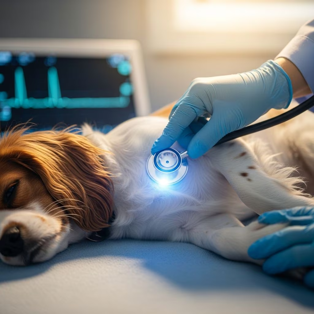
Atrial standstill represents a uncommon cardiac conduction abnormality where the atria fail to generate electrical impulses, leading to absent P waves on electrocardiograms. This condition disrupts normal heart rhythm, primarily manifesting as bradyarrhythmia in companion animals. Dogs, particularly certain breeds, and occasionally cats are affected, with presentations ranging from transient episodes to permanent states requiring intervention.
Understanding the Physiology of Atrial Standstill
The heart’s atria are crucial for coordinating blood flow into the ventricles. In normal sinus rhythm, P waves signify atrial depolarization. During atrial standstill, these waves vanish, and ventricular activity relies on escape rhythms, often resulting in slowed heart rates. This can be temporary, linked to electrolyte imbalances like hyperkalemia, or persistent, stemming from structural atrial pathology such as fibrosis or cardiomyopathy.
Temporary forms often resolve with correction of underlying issues, whereas persistent cases indicate intrinsic atrial disease. Research highlights atrial paralysis or standstill as key features, with histopathological findings revealing replacement fibrosis in affected tissues.
Common Causes and Risk Factors
Several etiologies contribute to this disorder. Hyperkalemia, frequently from urinary obstructions in cats or hypoadrenocorticism in dogs, triggers reversible standstill. Drugs like quinidine or digitalis, and myocardial injuries, can also induce transient episodes.
- Electrolyte disturbances: Elevated potassium levels suppress atrial excitability.
- Endocrine disorders: Addison’s disease in dogs leads to potassium retention.
- Structural heart changes: Idiopathic fibrosis, particularly in the right atrium, causes permanent standstill.
- Breed predispositions: English Springer Spaniels in dogs; Siamese cats show higher incidence.
- Other factors: Renal failure, toxicity, or chronic atrial dilation.
Persistent atrial standstill (PAS) often occurs in young adult dogs without identifiable metabolic causes, suggesting genetic or primary myocardial involvement.
Recognizing Clinical Signs in Dogs and Cats
Symptoms arise from reduced cardiac output and bradyarrhythmia. Owners frequently notice syncope, the most common presentation in PAS-affected dogs. Other signs include:
- Exercise intolerance and weakness.
- Fatigue and lethargy.
- Abdominal distension from ascites in right-sided heart failure.
- Muscle wasting, especially in forelimbs and shoulders.
- Collapse episodes mimicking seizures.
In cats, presentations may involve chylothorax or pleural effusions alongside bradycardia. Chronic cases can progress to congestive heart failure, with elevated creatinine signaling concurrent renal issues.
| Species | Primary Signs | Associated Findings |
|---|---|---|
| Dogs | Syncope, weakness | Ascites, muscle wasting |
| Cats | Pleural effusion, lethargy | Biatrial dilation |
Diagnostic Approaches for Accurate Identification
Diagnosis hinges on electrocardiography (ECG), revealing absent P waves and junctional or ventricular escape rhythms at rates of 40-60 bpm. Atropine response testing confirms persistence if no atrial activity resumes.
Additional tests include:
- Bloodwork: Serum electrolytes, renal parameters, and Addison’s screening.
- Echocardiography: Assesses atrial enlargement, ventricular function, and excludes structural diseases.
- Intracardiac electrograms: Verify absent atrial signals and pacing unresponsiveness.
- Thoracic imaging: Detects effusions or cardiomegaly.
Hyperkalemia or dehydration warrants immediate fluid therapy. Persistent cases show normal electrolytes and negative atropine tests.
Treatment Strategies: From Medical Management to Pacemakers
Management varies by type. Temporary standstill resolves with hyperkalemia correction via IV fluids and addressing causes like urinary obstruction.
For PAS, pacemaker implantation is definitive, restoring adequate heart rates and alleviating syncope. Studies report median survival of 866 days post-pacemaker, with favorable long-term outcomes.
- Medical therapy: Diuretics (furosemide), ACE inhibitors (enalapril), and low-sodium diets for heart failure.
- Pacemaker types: Single-chamber ventricular pacemakers suit most cases.
- Supportive care: Rest, monitoring for azotemia.
Post-implantation, syncope resolves, though cardiac remodeling like atrial dilation may persist. Regular ECG follow-ups ensure device efficacy.
Prognosis and Long-Term Outlook
Prognosis excels with prompt intervention. Pacemaker recipients show good survival, uninfluenced by initial congestive status or chamber sizes. Untreated cases, like a reported dog surviving four years on medical therapy alone, succumb to comorbidities such as renal azotemia.
Cats face guarded prognoses due to rarer occurrences and complications like restrictive pleural disease. Factors improving outcomes include early diagnosis, pacemaker placement, and comorbidity management.
Preventive Measures and Home Care Tips
While idiopathic PAS defies prevention, routine wellness checks aid early detection in predisposed breeds. Home care post-diagnosis involves:
- Stress-free environments to minimize exertion.
- Medication adherence and diet control.
- Observing for syncope recurrence or worsening ascites.
- Scheduled veterinary visits for pacemaker checks.
Owners should monitor appetite, activity, and abdominal girth, reporting changes promptly.
Frequently Asked Questions (FAQs)
What is the main difference between temporary and persistent atrial standstill?
Temporary resolves with electrolyte correction; persistent requires pacemakers due to atrial fibrosis.
Which dog breeds are most at risk?
English Springer Spaniels top the list, alongside mixed breeds.
Can cats recover without surgery?
Rarely; structural changes often necessitate advanced care, though medical management helps short-term.
How effective are pacemakers in pets?
Highly effective for symptom relief and extending survival to over two years median.
Is atrial standstill hereditary?
Possible in predisposed breeds, but research continues on genetic links.
Advances in Veterinary Cardiology Research
Recent studies emphasize PAS in young dogs, with pacemakers yielding positive remodeling despite persistent atrial issues. Histopathology confirms fibrosis as a hallmark, guiding future therapies. Ongoing work explores neurohormonal roles and ANP levels in progression.
References
- Clinical presentation, management, and survival in dogs with persistent atrial standstill — Journal of Veterinary Cardiology (Elsevier). 2024. https://pubmed.ncbi.nlm.nih.gov/38150959/
- Four-Year Follow-Up Persistent Atrial Standstill in a Dog — Israel Journal of Veterinary Medicine. 2019-06. http://www.ijvm.org.il/sites/default/files/correa_salgado.pdf
- Heart Beat Problems (Standstill) in Dogs — PetMD. N/A. https://www.petmd.com/dog/conditions/cardiovascular/c_dg_atrial_standstill
- Persistent atrial standstill in a cat — Journal of Veterinary Internal Medicine (PubMed). 1999. https://pubmed.ncbi.nlm.nih.gov/10561791/
- Atrial Standstill — Apple Valley Animal Hospital. N/A. https://www.applevalley.vet/sites/site-4271/documents/Atrial%20Standstill.pdf



