Lung Infection from Inhaled Materials in Pets
Understanding symptoms, diagnosis, and recovery from inhaled foreign material complications
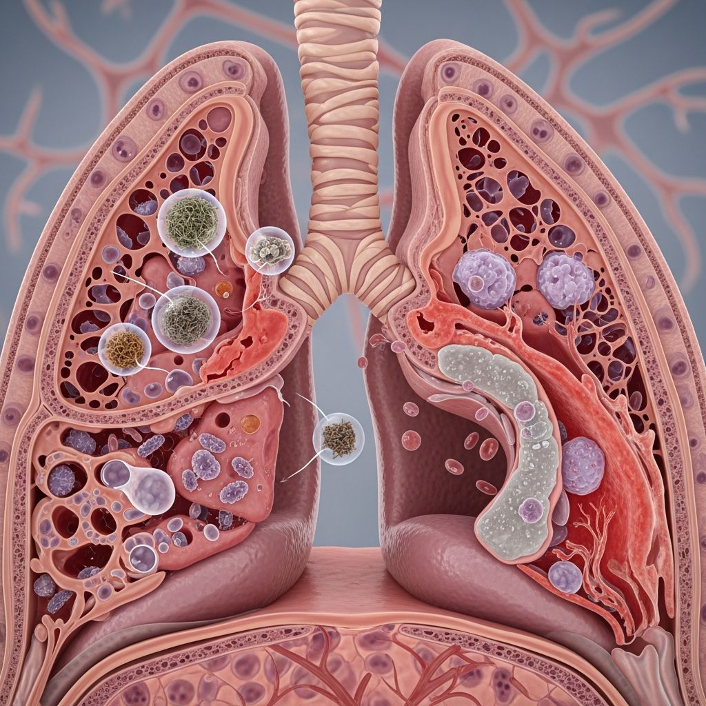
Aspiration pneumonia represents a serious respiratory condition that develops when pets accidentally inhale foreign substances into their lungs, such as vomit, liquid medications, food particles, or other materials. This inadvertent inhalation triggers inflammation within the lung tissue and creates an environment susceptible to bacterial infection, potentially compromising the animal’s ability to oxygenate blood effectively. Understanding this condition is essential for pet owners, as prompt recognition and veterinary intervention significantly improve outcomes.
Defining the Condition and Its Mechanism
Aspiration pneumonia occurs when substances that should remain in the digestive tract or oral cavity enter the respiratory tract instead. Unlike typical pneumonia caused by airborne pathogens, this condition develops through physical inhalation of material. The foreign substances irritate lung tissue, triggering an inflammatory cascade that damages the delicate structures responsible for gas exchange. Secondary bacterial colonization frequently occurs as the immune system struggles to manage the inflammatory insult, compounding the infection severity.
The condition affects both canine and feline patients, though specific epidemiological differences exist between species. Cats affected by this condition typically weigh around 3.8 kg on average, with equal gender distribution among affected individuals. The inflammatory response initiated by aspirated material predisposes the lungs to opportunistic bacterial infection, making antibiotic therapy a cornerstone of treatment protocols.
Recognizing Clinical Manifestations Across Species
Pet owners should remain vigilant for several clinical indicators that may suggest aspiration pneumonia. The presentation varies from subtle to severe, and early recognition dramatically improves treatment outcomes.
Common Signs in Cats
Feline patients typically present with a constellation of respiratory and systemic symptoms. Coughing, difficulty breathing, and fever represent the most frequently observed clinical signs. Additional indicators include decreased appetite, lethargy, nasal discharge, and respiratory distress. Some cats may exhibit less typical signs such as drooling, oral bleeding, or gastrointestinal symptoms including diarrhea.
Manifestations in Dogs
Canine patients demonstrate similar core symptoms but may present with additional clinical features. Coughing, respiratory distress, vomiting, and regurgitation constitute primary presenting complaints. Dogs may also exhibit exercise intolerance, fever, nasal discharge, difficulty swallowing, and increased respiratory effort that includes abdominal component involvement. In severe cases, blue-tinged gums or tongue (cyanosis) indicate critical oxygen deprivation requiring emergency intervention.
Subtle Presentations
Mild cases occasionally resemble “walking pneumonia” in humans, with affected pets showing minimal obvious symptoms. These animals may display only reduced appetite, mild lethargy, or decreased interest in normal activities such as following owners or engaging with toys. Such subtle presentations often go unrecognized, delaying necessary treatment, which underscores the importance of veterinary consultation when behavioral changes occur.
Diagnostic Approach and Imaging Techniques
Veterinary diagnosis requires systematic evaluation combining physical examination, laboratory assessment, and advanced imaging. The diagnostic process aims to confirm the presence of aspirated material, identify concurrent infections, and assess lung damage severity.
Physical Examination and Initial Assessment
Veterinarians begin with thorough physical examination, assessing respiratory effort, heart rate, temperature, and oxygen saturation. Fever, reduced oxygen levels, and abnormal white blood cell counts provide valuable diagnostic clues. Auscultation of lung fields may reveal crackling sounds or other abnormal breath sounds indicative of inflammation and fluid accumulation.
Imaging and Laboratory Diagnostics
Chest radiographs (X-rays) represent the primary diagnostic imaging modality, revealing patchy areas of inflammation or consolidation within lung tissue. These images may also document esophageal abnormalities such as megaesophagus, which increases aspiration risk. Bronchoalveolar lavage procedures involve infusing saline into airways, then retrieving samples via suction to collect cells for laboratory analysis. This technique identifies causative bacteria and guides targeted antibiotic selection.
Bloodwork assessment documents white blood cell changes and systemic inflammatory markers, while ultrasound examination can identify patchy lung lesions. In rare instances, additional diagnostics may include esophageal imaging to identify predisposing anatomical abnormalities.
Comprehensive Treatment Strategies
Treatment protocols focus on three primary objectives: managing inflammation, eliminating bacterial infection, and maintaining adequate oxygenation while supporting overall patient stability. The approach varies based on disease severity, underlying health conditions, and individual patient response.
Antibiotic Therapy
Antibiotics form the foundation of aspiration pneumonia treatment, either preventing secondary bacterial infection or treating existing infection. Common choices include Clavamox and doxycycline, typically administered for approximately two weeks. In hospitalized patients, intravenous antibiotic administration may achieve superior lung tissue penetration compared to oral formulations. The selection of specific antibiotics often depends on bronchoalveolar lavage culture results identifying causative organisms.
Oxygen Supplementation and Respiratory Support
Oxygen therapy becomes essential for patients with moderate to severe hypoxemia, defined as oxygen saturation below 94% or partial pressure of oxygen below 80 mm Hg on room air. Supplemental oxygen at 40-60% concentration continues until respiratory difficulty resolves and patients can transition to room air breathing. More than half of hospitalized cats require oxygen cage support, with duration varying from less than 24 hours to 96 hours depending on severity.
Animals demonstrating refractory pneumonia unresponsive to supplemental oxygen may require mechanical ventilation in intensive care settings to prevent ventilatory fatigue and potential respiratory collapse.
Supportive Care Modalities
Beyond antibiotics and oxygen, several adjunctive therapies optimize recovery outcomes:
- Nebulization delivers aerosolized medications and moisture to airways, facilitating clearance of secretions and reducing airway irritation
- Coupage (chest percussion) mechanically assists fluid mobilization from dependent lung regions, enhancing drainage and clearance
- Intravenous fluid therapy maintains hydration and supports blood pressure during acute illness phases
- Mucolytic agents such as N-acetylcysteine reduce respiratory secretion viscosity, particularly beneficial for animals with moderate to severe bronchiectasis or recurrent pneumonia
- Bronchodilators such as terbutaline open constricted airways, improving ventilation efficiency
- Antiemetic medications including Cerenia and ondansetron prevent vomiting, reducing aspiration risk during recovery
- Acid-reducing agents such as omeprazole decrease gastric reflux, minimizing future aspiration likelihood
Hospitalization and Monitoring
Severe cases require 24-hour facility hospitalization for continuous monitoring and intervention capability. Hospitalization duration varies considerably; one study documented median hospitalization of three days for affected cats, with ranges from zero to 21 days. Most cats recover after three days of hospitalization, though some require up to three weeks of inpatient care. Dogs typically require treatment spanning two to eight weeks depending on severity and underlying causes. During hospitalization, frequent vital sign monitoring, oxygen saturation assessment, and repeated imaging ensure treatment efficacy and permit timely intervention adjustments.
Recovery Trajectory and Long-term Management
Recovery timelines vary substantially based on disease severity, patient age, and concurrent health conditions. Mild cases may resolve in as little as three days, while severe presentations or animals with underlying health issues may require several months for complete resolution.
Post-Hospitalization Care
Discharged animals require continuation of prescribed antibiotics, typically completing approximately two-week courses. Environmental modifications enhance recovery by providing quiet, private resting spaces with humidifiers to moisten airways and facilitate secretion clearance. Owners should monitor for respiratory symptom changes or return of clinical signs necessitating immediate veterinary re-evaluation.
Follow-up Assessment
Veterinary rechecks following antibiotic completion include chest radiography to document pneumonia resolution. Additional antibiotics may be prescribed if radiographic evidence suggests incomplete resolution. Frequent rechecks continue until pneumonia completely resolves, ensuring treatment success and stable patient condition maintenance.
Prognosis and Outcome Expectations
Overall prognosis for aspiration pneumonia varies based on disease severity and promptness of treatment initiation. Recent research indicates encouraging survival rates; one study documented 89% survival for cats diagnosed with aspiration pneumonia. Prognosis ranges from fair to good overall, with many cases receiving prompt treatment demonstrating successful recovery.
However, severe cases involving inhalation of highly irritating substances or animals developing secondary bacterial infection face poorer prognosis. In rare instances, lung abscess formation may develop, necessitating additional therapeutic intervention. Advanced age and concurrent health conditions further compromise recovery likelihood and extend treatment timelines.
Risk Factors and Prevention Considerations
Certain conditions increase aspiration pneumonia susceptibility. Megaesophagus, a condition causing esophageal dilation, significantly elevates aspiration risk in both species. Animals requiring anesthesia demonstrate increased acid reflux events, though controlled studies assessing preventive antacid therapy effectiveness remain limited. Neurological conditions affecting swallowing, prolonged recumbency, and difficulties in positioning during feeding all contribute to aspiration risk.
Prevention strategies include careful medication administration techniques avoiding aspiration, appropriate feeding positioning, and prompt vomitus removal. Animals demonstrating regurgitation or swallowing difficulties warrant veterinary investigation to identify and address underlying causes before complications develop.
Frequently Asked Questions
How quickly does aspiration pneumonia develop after inhalation occurs?
Clinical signs may appear within hours to days following aspiration, though mild cases sometimes remain asymptomatic initially, resembling walking pneumonia in humans. The inflammatory response and secondary infection timeline varies with aspirated material type and volume.
Can aspiration pneumonia be prevented entirely?
Complete prevention is impossible, but risk reduction strategies include careful medication administration, proper feeding positioning, addressing underlying swallowing disorders, and prompt veterinary care for animals demonstrating regurgitation or aspiration risk factors.
Is aspiration pneumonia contagious between pets?
Aspiration pneumonia itself is not contagious, as it results from physical inhalation of material rather than infectious pathogen transmission. However, bacteria colonizing affected lungs could theoretically transmit to other animals through respiratory secretions, though this represents uncommon transmission.
What distinguishes aspiration pneumonia from infectious pneumonia?
Aspiration pneumonia develops from inhaled foreign material causing inflammation and secondary infection, while infectious pneumonia results from airborne pathogen inhalation. Diagnostic imaging, clinical history, and laboratory findings help differentiate between conditions.
When Emergency Care Becomes Necessary
Pet owners should seek immediate emergency veterinary attention when animals exhibit severe respiratory distress, blue-tinged gums or tongue, extreme lethargy, or inability to breathe effectively. These signs indicate critical oxygen deprivation requiring urgent intervention to prevent life-threatening deterioration. Even milder signs warrant prompt veterinary consultation, as early treatment substantially improves outcomes compared to delayed intervention.
References
- Aspiration Pneumonia in Cats – Causes, Treatment and Recovery — Vetster. 2024. https://vetster.com/en/conditions/cat/aspiration-pneumonia
- Aspiration Pneumonia in Cats: Causes and Treatment — PetMD. 2024. https://www.petmd.com/cat/conditions/respiratory/aspiration-pneumonia-in-cats
- Bacterial Pneumonia in Dogs and Cats: An Update — National Center for Biotechnology Information (NIH). 2020. https://pmc.ncbi.nlm.nih.gov/articles/PMC7114575/
- Aspiration Pneumonia in Cats – VETgirl Veterinary Continuing Education — VETgirl. 2023. https://vetgirlontherun.com/podcasts/aspiration-pneumonia-in-cats-vetgirl-veterinary-continuing-education-podcasts/
- Aspiration Pneumonia in Dogs — PetMD. 2024. https://www.petmd.com/dog/conditions/respiratory/aspiration-pneumonia-dogs
- What To Know About Aspiration Pneumonia In Pets — Texas A&M College of Veterinary Medicine & Biomedical Sciences. 2024. https://vetmed.tamu.edu/news/pet-talk/aspiration-pneumonia-in-pets/
- Aspiration Pneumonia in Dogs: Emergency Symptoms Explained — Garden State Veterinary Specialists. 2024. https://www.gsvs.org/woodbridge-nj/blog/dog-aspiration-pneumonia-emergency-symptoms/
Similar Articles







Read full bio of medha deb