

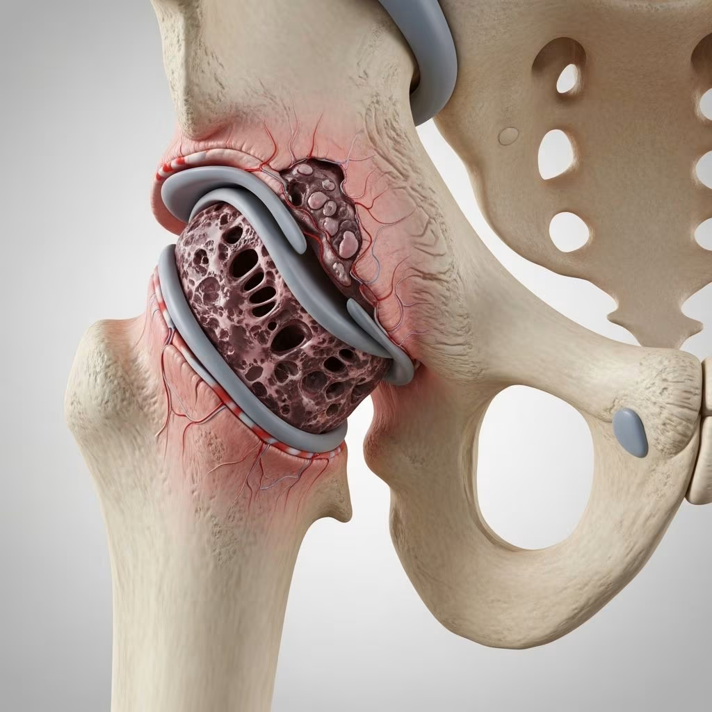
This orthopedic condition, often called Legg-Calvé-Perthes disease, strikes young small-breed dogs, leading to painful hip joint degeneration due to disrupted blood flow. Early detection and intervention are crucial for restoring mobility and preventing chronic arthritis.
Understanding the Condition
Aseptic necrosis involves the death of bone tissue in the femoral head without bacterial infection, primarily from vascular compromise. The femoral head, the ball-shaped top of the thigh bone that fits into the pelvis socket, loses its blood supply, causing bone collapse and joint instability. This self-limiting developmental disorder typically manifests in puppies under one year, though symptoms may appear later.
Unlike traumatic injuries, this is a non-inflammatory process where bone cells starve, leading to structural failure. Over time, the body attempts repair through granulation tissue, but secondary osteoarthritis often develops due to irregular joint surfaces.
Breeds and Age Groups at Risk
Small breeds dominate cases, with terriers, miniature poodles, Yorkshire terriers, and miniature pinschers showing high susceptibility. Studies note 60% of affected dogs are Yorkshire terriers, linked to genetic factors like autosomal recessive inheritance in some lines.
- High-risk breeds: Yorkshire Terrier, Miniature Pinscher, Terrier varieties, Poodle (miniature), Manchester Terrier
- Age onset: 4-11 months, averaging 5-7 months; rarely bilateral (10-17% of cases)
- Sex distribution: Equal in males and females
Genetic predisposition drives etiology, with multifactorial patterns interrupting endosteal blood supply to the epiphysis and metaphysis.
Recognizing Clinical Symptoms
Owners often notice gradual hindlimb lameness, starting subtly and worsening over weeks. Pain intensifies on hip manipulation, especially extension or abduction, prompting behavioral changes like irritability or reluctance to play.
| Symptom | Description | Frequency |
|---|---|---|
| Hindlimb lameness | Progressive, unilateral (most common) | Common |
| Pain on hip exam | Especially abduction/extension | High (e.g., 28/35 cases) |
| Muscle atrophy | Wasting, greater trochanter prominence | Frequent (25/35 cases) |
| Limb shortening | Apparent due to collapse | Moderate (14/35 cases) |
| Crepitus | Grating during movement | Occasional (8/35 cases) |
Advanced signs include non-weight-bearing, stair avoidance, and medial patellar luxation comorbidity.
Pathophysiological Mechanisms
The process begins with ischemia: blood vessels supplying the femoral head infarct, triggering epiphyseal and metaphyseal necrosis. Trabecular bone collapses, fracturing subchondral support and cracking overlying cartilage. This incongruity sparks instability, inflammation, and eventual degenerative joint disease.
Repair involves vascular ingrowth replacing necrotic bone via creeping substitution, but remodeling often yields a deformed head, perpetuating pain. Concurrent issues like patellar luxation exacerbate lameness.
Diagnostic Approaches
Veterinarians rely on history, physical exam, and imaging. Palpation reveals pain, reduced range of motion, atrophy, and crepitus. Radiographs confirm diagnosis, showing:
- Flattening or collapse of femoral head
- Moth-eaten or irregular density
- Neck sclerosis or deformity
- Joint space widening, osteophytes
Early films may appear normal; serial imaging tracks progression. Advanced cases show osteoarthritis.
Treatment Strategies
Conservative management suits pre-collapse cases: strict rest via sling or crate, NSAIDs for pain, and physiotherapy. Only 25% respond fully, with arthritis risk rising. Surgery is gold standard post-collapse.
Femoral Head and Neck Ostectomy (FHO): Removes necrotic head/neck, allowing scar tissue and muscles to form a pseudo-joint. Ideal for small dogs (<12kg), yielding excellent function. Total hip replacement suits larger breeds or bilateral disease.
- Conservative: Exercise restriction, analgesics; monitor 4 weeks
- Surgical: FHO (preferred); THR if needed
Postoperative Care and Recovery
Post-FHO, patients receive antibiotics (e.g., cephalexin), analgesics (tramadol, buprenorphine), and passive range-of-motion exercises. Physiotherapy accelerates healing: underwater treadmill, laser therapy. Most bear weight within days; full recovery spans 3-6 months with follow-ups.
Prognosis excels: 90%+ achieve good limb use, though mild intermittent lameness may persist. Early intervention minimizes arthritis.
Preventive Measures and Genetic Considerations
No cure exists for genetic predisposition, but screening breeding stock reduces incidence. Avoid over-exercise in at-risk puppies. Routine hip checks aid early detection.
Long-Term Outcomes and Complications
FHO boasts high success: studies show sustained mobility years post-op. Complications include infection (<5%), delayed healing, or nerve damage—rare with skilled surgeons. Bilateral cases may need staged procedures.
Frequently Asked Questions (FAQs)
What causes aseptic femoral head necrosis in dogs?
Primarily genetic vascular disruption leading to bone death; exact trigger unknown but breed-linked.
Can my dog recover without surgery?
Possible in early stages with rest (25% success), but surgery recommended for reliable outcomes.
How long does FHO recovery take?
Weight-bearing in 1-2 weeks; full function in 3-6 months with rehab.
Is this condition painful for dogs?
Yes, causing significant lameness and discomfort until treated.
Which breeds need hip screening?
Terriers, Poodles, Miniature Pinschers, Yorkshire Terriers—especially juveniles.
Owner Tips for Managing Affected Dogs
- Monitor for limping early
- Maintain ideal weight to reduce joint stress
- Follow rehab protocols diligently
- Consider bilateral screening via radiographs
References
- Hip: aseptic femoral head/neck necrosis in Dogs (Canis) — Vetlexicon. Accessed 2026. https://www.vetlexicon.com/canis/musculoskeletal/articles/hip-aseptic-femoral-headneck-necrosis/
- Long-term follow-up of avascular necrosis of the femoral head — PubMed (de CJ Piek et al.). 1996-06-01. https://pubmed.ncbi.nlm.nih.gov/8642794/
- Avascular Necrosis of Femoral Head in Pets — Monvet. Accessed 2026. https://monvet.com/en/femoral-head-necrosis-pets/
- Aseptic Femoral Head Necrosis in Dogs — WagWalking. Accessed 2026. https://wagwalking.com/condition/aseptic-femoral-head-necrosis
- Legg-Calve-Perthes Disease — VCA Animal Hospitals. Accessed 2026. https://vcahospitals.com/know-your-pet/necrosis-of-the-femoral-head-in-dogs
- Legg-Calvé-Perthes Disease in the Dog: A Case-Based Review — SAVA Vetnews. 2023-03. https://www.sava.co.za/vetnews/2016/2023%20March/VN%20CPD%20March%202023.pdf



