

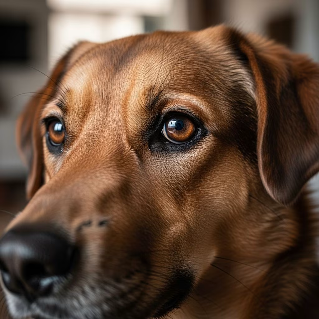
Understanding Anisocoria in Dogs
Anisocoria is a condition characterized by unequal pupil sizes in dogs, where one pupil appears noticeably larger or smaller than the other. This asymmetry in pupil size can be a sign of various underlying health conditions ranging from minor eye irritations to serious neurological disorders. Pet owners who notice this symptom should seek veterinary attention promptly, as early diagnosis and treatment can help prevent complications and protect their dog’s vision. Understanding what anisocoria is, its potential causes, and available treatment options is essential for responsible pet care.
What Causes Anisocoria in Dogs?
Anisocoria can result from multiple different conditions affecting various systems in a dog’s body. The causes typically fall into several categories, including ocular (eye-related) problems, neurological disorders, and autonomic nervous system dysfunction.
Ocular Causes
Eye trauma represents one of the most common causes of anisocoria in dogs. Damage to the cornea, such as corneal ulcers, can cause one pupil to react differently than the other. Uveitis, which is inflammation within the eye, often causes one pupil to remain constricted while the other maintains normal size. This occurs because prostaglandins released during inflammation directly affect the iris sphincter muscle.
Glaucoma, characterized by elevated pressure within the eye, can cause the affected pupil to remain permanently dilated. Additionally, iris degeneration, which is particularly common in older dogs, occurs when the muscles within the iris weaken and no longer function properly, resulting in unequal pupil sizes. Posterior synechia, an adherence of the iris to the lens, can also produce anisocoria through various mechanisms including miosis, mydriasis, or dyscoria.
Neoplasia of the iris and ciliary body can cause anisocoria through multiple pathways, including prostaglandin-mediated uveitis leading to miosis, secondary glaucoma causing mydriasis, synechia formation, or direct mass infiltration of the iris with physical obstruction of the pupil.
Neurological Causes
Horner’s syndrome is a significant neurological condition that affects the nerves controlling eye muscles and can result in anisocoria. This syndrome involves dysfunction of the sympathetic nervous system and may be associated with otitis media, which accounts for 1% to 4% of sympathetic dysfunction cases in dogs.
Head trauma is a common cause of parasympathetic dysfunction, resulting from compression of the midbrain at the level of the parasympathetic nuclei of cranial nerve III. This compression typically occurs secondary to hemorrhage or increased intracranial pressure, resulting in herniation of the brain tissue.
Central nervous system diseases, whether immune-mediated, infectious, or neoplastic, can cause visual, sympathetic, or parasympathetic dysfunction in various combinations. Infectious agents implicated in CNS disease include viruses, bacteria, fungi, protozoa, and rarely rickettsial infections.
Iatrogenic Causes
Anisocoria can develop as a result of medical procedures themselves. Jugular venipuncture can cause anisocoria due to damage to the second-order sympathetic neuron as it courses through the jugular groove, deep to the jugular vein. Excessive probing for the vein during blood draws may result in damage to surrounding structures.
Recognizing Symptoms of Anisocoria
The primary symptom of anisocoria is the visible difference in pupil sizes between the two eyes. However, dogs with anisocoria may exhibit additional symptoms depending on the underlying cause:
- Excessive blinking or blepharospasm
- Eye redness and hyperemia
- Corneal cloudiness or edema
- Aqueous flare within the eye
- Sensitivity to light (photophobia)
- Discharge from the affected eye
- Behavioral changes related to vision impairment
- Signs of pain or discomfort
If your dog suddenly develops unequal pupils, immediate veterinary care is warranted to minimize the risk of blindness and other serious complications.
Diagnostic Process for Anisocoria
Veterinarians employ several diagnostic methods to identify the cause of anisocoria and localize the lesion within the nervous or visual system.
Physical Examination
The initial evaluation includes a thorough ophthalmologic examination to assess pupil response to light, size, and shape. The veterinarian will examine both eyes for signs of inflammation, discharge, corneal damage, or other visible abnormalities. Assessment of iris appearance, clarity of the lens, and overall eye health is essential before investigating neurological causes.
Pharmacologic Testing
Pharmacologic testing can aid practitioners with lesion localization within the sympathetic or parasympathetic system.
Physostigmine Testing: Physostigmine (0.5%) is a cholinesterase inhibitor that requires an intact postganglionic neuron to induce miosis. Topical administration differentiates between preganglionic and postganglionic parasympathetic lesions. When administered to both eyes, if a preganglionic lesion is present, it causes rapid pupil constriction relative to the normal eye. In a normal eye, administration causes slow or delayed constriction.
Pilocarpine Testing: Dilute pilocarpine (0.2%-1% solution) is a parasympathomimetic alkaloid used for diagnostic purposes. Animals with ocular parasympathetic dysfunction demonstrate rapid pupillary constriction following pilocarpine administration, while those with iris atrophy show only partial constriction. Posterior synechia results in no or minimal response to pilocarpine.
Phenylephrine Testing: Dilute phenylephrine (1%) can be used to test the sympathetic system. When applied topically to both eyes, it should cause rapid dilation (typically within 20 minutes) in an eye with postganglionic sympathetic neuron dysfunction, while administration in a normal eye produces no effect.
Additional Diagnostic Tests
Depending on the suspected cause, veterinarians may recommend imaging studies such as MRI or CT scans to evaluate the brain and nervous system, blood work to assess for systemic infections or immune-mediated diseases, or specialized ophthalmologic testing including tonometry to measure intraocular pressure and assess for glaucoma.
Treatment Options for Anisocoria
The treatment of anisocoria depends entirely on the underlying cause of the condition, and specific treatment will be tailored specifically to the diagnosis. Treatment approaches can range from no recommended intervention to surgical procedures.
Medical Treatments
Many conditions causing anisocoria can be effectively managed with medications:
- Antibiotic or antifungal eye drops: Used for infections or corneal ulcers to eliminate pathogens and promote healing
- Pain relief medications: Help manage discomfort and inflammation associated with eye conditions
- Glaucoma medications: Lower intraocular pressure to prevent further damage and vision loss
- Anti-inflammatory drugs: Reduce swelling from uveitis, neurological conditions, or other inflammatory processes
- Immunosuppressive therapy: Required for immune-mediated CNS disease, though infectious and neoplastic causes must first be eliminated
Surgical Interventions
Surgical treatment becomes necessary for severe cases:
- Severe eye injuries requiring repair or reconstruction
- Cataracts affecting vision
- Tumors or neoplastic growths requiring removal
- Advanced glaucoma cases unresponsive to medical management
Long-term Management
Some conditions, such as corneal injuries or damage to sympathetic nerves, can be effectively treated with resolution of anisocoria. However, conditions like uveitis and glaucoma can only be managed long-term, and anisocoria may resolve with appropriate management of the underlying disease. If your dog is diagnosed with a chronic condition like glaucoma, long-term management may be necessary to prevent future episodes of anisocoria or other complications.
Prognosis and Recovery
The prognosis for dogs with anisocoria varies significantly depending on the underlying cause, the severity of the condition, and how quickly treatment is initiated. Many cases of anisocoria caused by minor eye injuries or infections respond well to appropriate medical therapy. However, anisocoria resulting from serious neurological conditions or advanced eye diseases may have a more guarded prognosis. Early detection and prompt veterinary intervention significantly improve the likelihood of preserving vision and achieving a favorable outcome.
When to Seek Veterinary Care
Pet owners should contact a veterinarian immediately if they notice:
- Sudden development of unequal pupil sizes
- Significant change in pupil appearance or responsiveness
- Accompanying eye pain, redness, or discharge
- Vision changes or signs of blindness
- Head trauma or recent injury
- Neurological symptoms such as disorientation or seizures
Frequently Asked Questions About Anisocoria in Dogs
Q: Is anisocoria always a sign of a serious problem?
A: While anisocoria can indicate serious underlying conditions, some causes such as minor corneal injuries or temporary inflammation may resolve with appropriate treatment. However, it should always be evaluated by a veterinarian to determine the cause.
Q: Can anisocoria be prevented?
A: While not all cases can be prevented, you can reduce the risk by protecting your dog’s eyes from trauma, maintaining regular veterinary check-ups, promptly treating eye infections or inflammation, and avoiding exposure to harmful chemicals or irritants.
Q: How quickly should I seek veterinary care if I notice unequal pupils?
A: If anisocoria develops suddenly, immediate veterinary care is warranted to minimize the risk of blindness and complications. If the change is gradual, schedule an appointment as soon as possible, but seek emergency care if accompanied by other symptoms like eye pain or vision loss.
Q: Will my dog go blind from anisocoria?
A: Blindness is not an automatic consequence of anisocoria. The outcome depends on the underlying cause and how quickly treatment is initiated. Many conditions respond well to appropriate therapy, preserving vision.
Q: Can anisocoria resolve on its own?
A: Some cases may improve, but this depends on the underlying cause. Most cases require veterinary intervention and specific treatment directed at the cause to achieve resolution.
Q: What is the difference between anisocoria and other pupil abnormalities?
A: Anisocoria specifically refers to unequal pupil sizes. Other abnormalities might include fixed pupils (non-responsive to light), oval-shaped pupils, or abnormal dilation or constriction patterns.
Conclusion
Anisocoria in dogs requires prompt veterinary attention and appropriate diagnostic workup to identify the underlying cause. Whether the condition results from eye trauma, infection, inflammation, or neurological disease, early intervention provides the best opportunity for successful treatment and vision preservation. By understanding the potential causes, recognizing symptoms, and seeking timely veterinary care, pet owners can help ensure their dogs receive the necessary treatment to address this condition and maintain their quality of life.
References
- The Practitioner’s Guide to Neurologic Causes of Canine Anisocoria — Today’s Veterinary Practice. 2016-05. https://todaysveterinarypractice.com/ophthalmology/the-practitioners-guide-neurologic-causes-canine-anisocoria/
- Anisocoria in Dogs: Symptoms, Causes, Diagnosis, & Treatment — Muffin’s Halo. 2024. https://www.muffinshalo.com/blogs/news/anisocoria-in-dogs
- Anisocoria in Dogs: Causes & Treatment — Animal Emergency Urgent Care. 2024. https://www.animalemergencyurgentcare.com/anisocoria-in-dogs/
- Anisocoria in Dogs — VCA Animal Hospitals. 2024. https://vcahospitals.com/know-your-pet/anisocoria-in-dogs
- Anisocoria in Dogs – PetMD — PetMD. 2024. https://www.petmd.com/dog/conditions/eyes/anisocoria-dogs
- Unequal Pupils (Anisocoria) in Dogs – Causes, Treatment and Symptoms — Vetster. 2024. https://vetster.com/en/symptoms/dog/unequal-pupils-anisocoria
- Anisocoria in Dogs – Symptoms, Causes, Diagnosis, Treatment — Wag Walking. 2024. https://wagwalking.com/condition/anisocoria



