

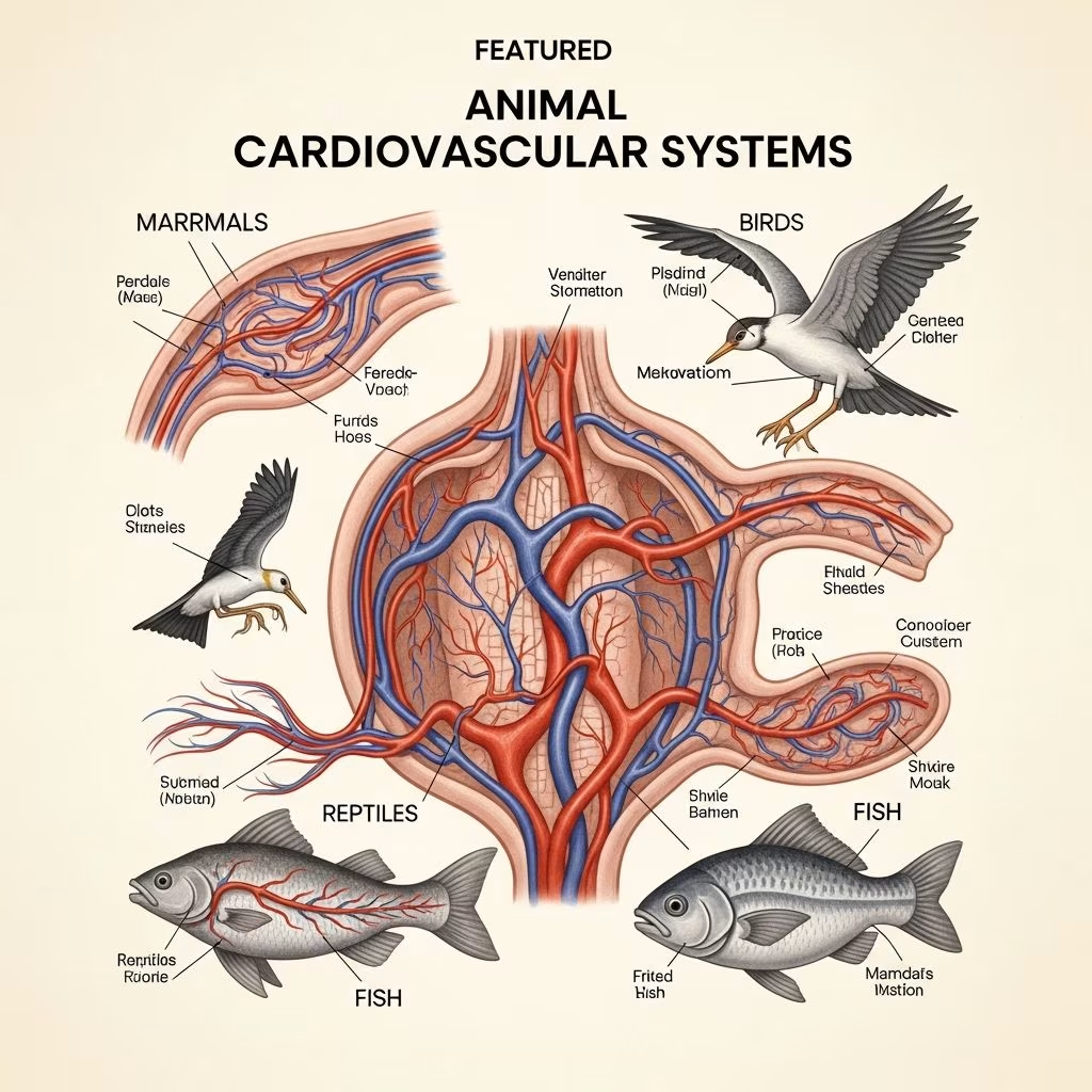
The cardiovascular system forms the lifeline of animals, transporting oxygen, nutrients, and waste through a network of the heart and blood vessels. This vital organ system varies across species to meet unique physiological demands, ensuring survival from fish to mammals.
Core Components of the Circulatory Network
At its foundation, the system includes the heart as the central pump, arteries carrying oxygenated blood away from it, veins returning deoxygenated blood, and capillaries facilitating exchange with tissues. In vertebrates, this setup creates a closed circuit where blood remains confined within vessels, enabling efficient unidirectional flow.
- Heart: A muscular organ with chambers that contract rhythmically to propel blood.
- Arteries: Thick-walled vessels distributing high-pressure blood from the heart.
- Veins: Thinner vessels with valves to prevent backflow, guiding blood back to the heart.
- Capillaries: Microscopic channels where gas and nutrient exchange occurs via diffusion.
This architecture supports double circulation in higher vertebrates: pulmonary for lung oxygenation and systemic for body distribution.
Heart Anatomy Across Animal Classes
Hearts evolve in complexity from two chambers in fish to four in mammals and birds. Fish possess a simple tube-like heart pumping blood sequentially through chambers: sinus venosus, atrium, ventricle, and conus arteriosus.
Amphibians advance to three chambers—two atria and one ventricle—allowing partial separation of oxygenated and deoxygenated blood, though mixing reduces efficiency.
| Species Group | Chamber Count | Circulation Type | Key Adaptation |
|---|---|---|---|
| Fish | 2 | Single | Sequential pumping |
| Amphibians | 3 | Double (partial) | Atrial separation |
| Reptiles | 3-4 | Double (variable) | Foramen of Panizza |
| Birds/Mammals | 4 | Double (complete) | Ventricular septum |
Reptiles, especially crocodilians, feature a four-chambered heart with adaptations like the foramen of Panizza—a shunt between ventricles enabling blood rerouting during dives, controlled by neural and hormonal signals.
Mammals and birds exhibit fully divided four-chambered hearts: right atrium/ventricle for pulmonary circuit, left for systemic. A thick interventricular septum prevents mixing, crucial for endothermy.
Electrical Conduction: Triggering the Beat
The heartbeat originates from specialized pacemaker cells. In mammals, the sinoatrial (SA) node in the right atrium initiates depolarization, spreading via internodal pathways to the atrioventricular (AV) node. This delay ensures atrial contraction precedes ventricular.
The impulse then races through the AV bundle (His bundle), bundle branches, and Purkinje fibers to ventricular myocardium, causing coordinated squeeze from apex upward.
Birds differ: depolarization starts subepicardially at the right ventricular apex, yielding distinct ECG patterns like negative QRS in lead II, akin to horses. Their larger Purkinje fibers and smaller cardiomyocytes support elevated heart rates.
- SA Node: Sets pace, influenced by autonomic nerves.
- AV Node: Delays for atrial emptying.
- Purkinje Network: Rapid ventricular spread.
Monotremes like platypus show branched right bundle branches extending to subendocardial networks.
Valvular Mechanisms for Unidirectional Flow
Four main valves maintain directionality: tricuspid and mitral (AV valves) between atria/ventricles; pulmonic and aortic (semilunar) at great vessel exits. Chordae tendineae and papillary muscles anchor AV valves, preventing prolapse.
Venous valves in larger veins combat gravity, especially in limbs. In dogs, the heart resides in the pericardial sac between ribs 3-6, with coronary arteries nourishing its muscle.
Contractile Machinery and Energy Dynamics
Cardiomyocytes rely on excitation-contraction coupling: depolarization opens calcium channels, triggering sarcoplasmic reticulum release for actin-myosin cross-bridging. ATP powers this and relaxation via reuptake.
Oxygen drives 95% of ATP production via mitochondria. Birds demand more due to flight, boasting double mammalian cardiac mass relative to size and higher outputs.
Circulatory Variations by Species
Mammalian systemic circulation delivers oxygenated blood via aorta branches; pulmonary via pulmonary arteries to lungs. Reptilian shunts like crocodilian conus ridges adjust flows for diving.
Endotherms’ complete separation sustains high metabolism; ectotherms tolerate mixing for energy conservation.
Pericardium and Coronary Nourishment
The pericardium encases the heart, providing lubrication and protection. Coronary arteries branch from the aortic root, ensuring constant supply despite compression during systole.
Physiological Demands and Adaptations
Cardiac output scales with body size and activity: small mammals have rapid rates (e.g., shrews >1000 bpm), large ones slower. Birds’ fast sinus rates match aerial demands.
Lymphatics complement by returning interstitial fluid, preventing edema.
Maintaining Cardiovascular Health in Animals
Veterinary monitoring involves auscultation, ECG, echocardiography. Common issues like murmurs signal valve defects; arrhythmias indicate conduction flaws.
Frequently Asked Questions (FAQs)
What is the main difference between fish and mammalian hearts?
Fish hearts have two chambers with single circulation; mammals have four with complete double circulation separating oxygenated/deoxygenated blood.
How do birds’ hearts differ from mammals’?
Birds have higher cardiac mass, larger Purkinje fibers, and apex-initiated depolarization for faster rates supporting flight.
Why do crocodiles have unique heart features?
The foramen of Panizza and conus sphincter allow ventricular shunting during dives, bypassing lungs.
What powers heart contractions?
Calcium cycling and ATP from oxidative metabolism, with oxygen essential for most energy.
Where is the dog heart located?
Between right/left lungs in the pericardial sac, spanning ribs 3-6.
References
- The Cardiovascular System in Animals — Merck Veterinary Manual. 2023. https://www.merckvetmanual.com/circulatory-system/cardiovascular-system-introduction/the-cardiovascular-system-in-animals
- Structure and Function of the Cardiovascular System in Dogs — PetPlace (Veterinary Resource). 2022. https://www.petplace.com/article/dogs/pet-health/structure-and-function-of-the-cardiovascular-system-in-dogs
- The heart and heart conducting system in the kingdom of animals — PMC (Peer-reviewed). 2008-04-01. https://pmc.ncbi.nlm.nih.gov/articles/PMC2323757/
- Overview of the Circulatory System — University of Oregon (Academic). 2023. https://opentext.uoregon.edu/animalphysiology/chapter/6-1-overview-of-the-circulatory-system/
- Animal Circulatory Systems — Georgia Tech Biosci (.edu). 2023. https://organismalbio.biosci.gatech.edu/nutrition-transport-and-homeostasis/animal-circulatory-systems/
- Animal Organs: Cardiovascular System — University of Vigo (.edu equivalent). 2022. https://mmegias.webs.uvigo.es/02-english/2-organos-a/guiada_o_a_05cardiovascular.php



