

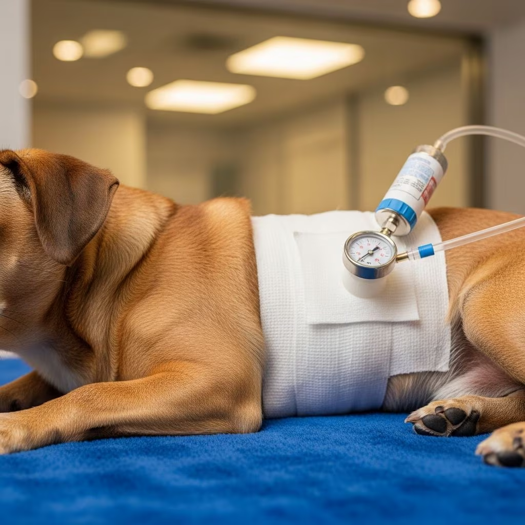
Negative pressure wound therapy (NPWT), often called vacuum-assisted closure, has transformed how veterinarians manage challenging injuries in dogs and cats. This technique applies controlled suction to wounds, promoting faster tissue repair, controlling infection risks, and preparing sites for surgery or natural closure.
Understanding the Core Principles of NPWT
At its heart, NPWT creates a sealed environment over the wound using specialized foam and an airtight drape. A connected pump generates subatmospheric pressure, typically between 90-125 mmHg, which draws out fluids while mechanically stimulating the healing tissues. This dual action not only removes excess exudate but also shrinks dead space, enhances blood flow, and accelerates granulation tissue formation.
The process begins with thorough wound cleaning and debridement to eliminate debris and necrotic material. A black polyurethane foam is then placed into the wound bed, extending slightly beyond the edges to ensure even pressure distribution. An adhesive drape seals the area, and tubing connects to the vacuum unit, which collects fluids in a disposable canister for monitoring.
- Fluid Management: Excess moisture is suctioned away, reducing edema and bacterial load.
- Tissue Perfusion: Micro-deformations from the vacuum improve circulation, delivering oxygen and nutrients.
- Granulation Boost: Healthy pink tissue grows rapidly, often within 3-4 days.
Key Benefits in Small Animal Practice
Clinical studies highlight NPWT’s superiority over traditional moist dressings. In dogs, wounds treated with vacuum therapy develop granulation beds in 3-4 days versus 6-7 days with wet-to-dry bandages. Cats show similar gains, with shorter times to closure compared to foam dressings alone.
| Benefit | Traditional Care | NPWT |
|---|---|---|
| Granulation Time | 6-7 days | 3-4 days |
| Dressing Changes | Daily | Every 3-4 days |
| Time to Reconstruction | 14 days | 7 days |
| Anesthesia Events | Multiple | Fewer |
These improvements translate to cost savings, less stress for patients, and better outcomes in trauma cases. NPWT also stabilizes surgical flaps and grafts, increasing take rates by minimizing shear and fluid buildup.
Ideal Scenarios for Application
NPWT shines in managing open traumatic wounds, such as bite injuries, vehicle accidents, or degloving trauma common in small animals. It’s particularly useful for deep or irregular defects where contraction aids closure.
- Contaminated or infected wounds after initial debridement.
- Post-surgical sites at risk of dehiscence.
- Large skin deficits on limbs, trunk, or tail.
- Preparation for skin grafts or flaps.
- Chronic non-healing ulcers in older pets.
In felines, where wounds often involve abscesses or fight-related punctures, NPWT facilitates outpatient care after initial hospital sessions, reducing costs and hospitalization time.
Step-by-Step Guide to Implementation
Successful NPWT requires meticulous preparation. Start with sedation or anesthesia for pain control and precise placement.
- Prep the Wound: Clip hair widely, aseptically clean, and sharply debride all devitalized tissue.
- Seal the Periphery: Apply skin protectant spray or ostomy paste around edges for better drape adhesion, especially on mobile areas like limbs.
- Place Foam: Cut foam to fit snugly, avoiding overpacking to prevent pressure points.
- Apply Drape: Ensure a complete airtight seal, trimming excess and smoothing wrinkles.
- Connect and Activate: Attach tubing, set to continuous -125 mmHg (adjust lower for sensitive patients), and confirm seal integrity.
- Monitor: Check canister exudate daily for changes in volume or color indicating infection.
Dressing changes occur every 3-4 days, allowing extended wear without frequent interventions. Transition to outpatient use once stable.
Species-Specific Considerations
Dogs: Robust Responders
Larger breeds with high-mobility wounds benefit from NPWT’s contraction effect in deep cavities. Studies confirm quicker revascularization and reduced edema in canine models.
Cats: Delicate but Effective
Feline skin is thinner and more prone to maceration, so lower pressures (80-100 mmHg) and frequent seal checks are advised. Retrospective data shows NPWT outperforms standard dressings for open management, though cat-specific trials are limited.
Potential Challenges and Avoidance Strategies
While effective, NPWT isn’t risk-free. Common issues include poor seals on curved surfaces, foam entrapment in healing tissue, or discomfort from continuous suction.
| Challenge | Solution |
|---|---|
| Seal Failure | Ostomy paste ring; skin prep spray. |
| Pain on Activation | |
| Foam Adherence | Cut precisely; change every 72 hours. |
| Exudate Overload | Smaller canister; frequent empties. |
Contraindications include untreated osteomyelitis, malignancy over the site, or exposed vessels/tendons without protection. Monitor for toxicity from foam breakdown products, though rare in vets.
Clinical Evidence and Outcomes
Veterinary adoption of NPWT grew from human trauma applications, now backed by small animal studies. A canine trial showed cost-neutral benefits with halved reconstruction times. Feline cases report full closure in under two weeks versus standard care. Overall, NPWT reduces complications like seroma and infection by 50% in some series.
Future directions include portable disposable units for first-opinion clinics and intermittent pressure protocols tailored to species.
Frequently Asked Questions (FAQs)
What pressure setting is best for small animals?
Start at -125 mmHg continuous for dogs; -75 to -100 mmHg for cats to minimize discomfort.
How often should dressings be changed?
Every 3-4 days, or sooner if seal fails or exudate fills canister.
Is NPWT safe for infected wounds?
Yes, after debridement; it reduces bacterial counts by continuous drainage.
Can I use NPWT at home?
Outpatient use is feasible after initial vet sessions, with training.
What are the costs involved?
Initial setup $200-500; comparable to multiple bandage changes long-term.
Integrating NPWT into Practice
For busy clinics, NPWT streamlines wound care by extending intervals and enabling earlier discharges. Training via workshops ensures proficiency. As evidence mounts, it promises to become standard for complex cases, improving pet welfare and owner satisfaction.
References
- Negative Pressure Wound Therapy (NPWT) in small animal medicine — Thieme Connect. 2015. https://www.thieme-connect.com/products/ejournals/pdf/10.15654/TPK-150957.pdf?articleLanguage=en
- Reviewing negative pressure wound therapy in animals — Vet Times. 2024-11-12. https://www.vettimes.com/news/vets/small-animal-vets/reviewing-negative-pressure-wound-therapy-in-animals
- Current concepts in negative pressure wound therapy — PubMed. 2015. https://pubmed.ncbi.nlm.nih.gov/25744142/
- Filling the vacuum: Role of negative pressure wound therapy in … — PubMed. 2021. https://pubmed.ncbi.nlm.nih.gov/34428942/
- Filling the vacuum: Role of negative pressure wound therapy in … — Sage Journals. 2021. https://journals.sagepub.com/doi/10.1177/1098612X211037873
- CVC highlight: Wound therapy: The positives of negative pressure — dvm360. N/A. https://www.dvm360.com/view/cvc-highlight-wound-therapy-positives-negative-pressure



